8IK7 | pdb_00008ik7

Cryo-EM structure of hnRAC1 fibril.


Experimental Data Snapshot

  • Method: ELECTRON MICROSCOPY
  • Resolution: 3.69 Å
  • Aggregation State: FILAMENT 
  • Reconstruction Method: HELICAL 

wwPDB Validation 3D Report Full Report

Validation slider image for 8IK7

This is version 1.0 of the entry. See complete history

Literature

Formed fibrils of hnRNP A1 reversible amyloid core GFGGNDNFG (residues 209-217)

Li, D.N.Ma, Y.Y.Li, D.Dai, B.Liu, C.

To be published.

Macromolecule Content 

  • Total Structure Weight: 21.21 kDa 
  • Atom Count: 1,488 
  • Modeled Residue Count: 216 
  • Deposited Residue Count: 216 
  • Unique protein chains: 1

Macromolecules

Find similar proteins by:  Sequence   |   3D Structure  
Entity ID: 1
MoleculeChains  Sequence LengthOrganismDetailsImage
GLY-PHE-GLY-GLY-ASN-ASP-ASN-PHE-GLY9Homo sapiensMutation(s): 0 
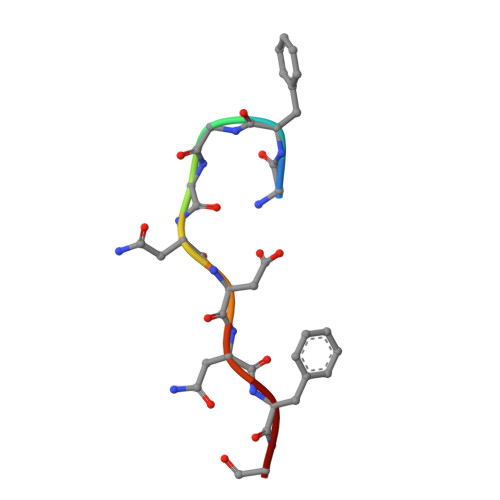
UniProt & NIH Common Fund Data Resources
Find proteins for P09651 (Homo sapiens)
Explore P09651 
Go to UniProtKB:  P09651
PHAROS:  P09651
GTEx:  ENSG00000135486 
Entity Groups
UniProt GroupP09651
Sequence Annotations
Expand
Reference Sequence

Experimental Data & Validation

Experimental Data

  • Method: ELECTRON MICROSCOPY
  • Resolution: 3.69 Å
  • Aggregation State: FILAMENT 
  • Reconstruction Method: HELICAL 

Structure Validation

View Full Validation Report


Entry History 

& Funding Information

Deposition Data


Funding OrganizationLocationGrant Number
Not funded--

Revision History  (Full details and data files)

  • Version 1.0: 2024-03-06
    Type: Initial release