8IB0 | pdb_00008ib0

The amyloid structure of mouse RIPK1 RHIM-containing domain by solid-state NMR


Experimental Data Snapshot

  • Method: SOLID-STATE NMR
  • Conformers Calculated: 196 
  • Conformers Submitted: 10 
  • Selection Criteria: structures with the lowest energy 

wwPDB Validation 3D Report Full Report

Validation slider image for 8IB0

This is version 1.3 of the entry. See complete history

Literature

The structure of mouse RIPK1 RHIM-containing domain as a homo-amyloid and in RIPK1/RIPK3 complex.

Liu, J.Wu, X.L.Zhang, J.Li, B.Wang, H.Y.Wang, J.Lu, J.X.

(2024) Nat Commun 15: 6975-6975

  • DOI: https://doi.org/10.1038/s41467-024-51303-y
  • Primary Citation Related Structures: 
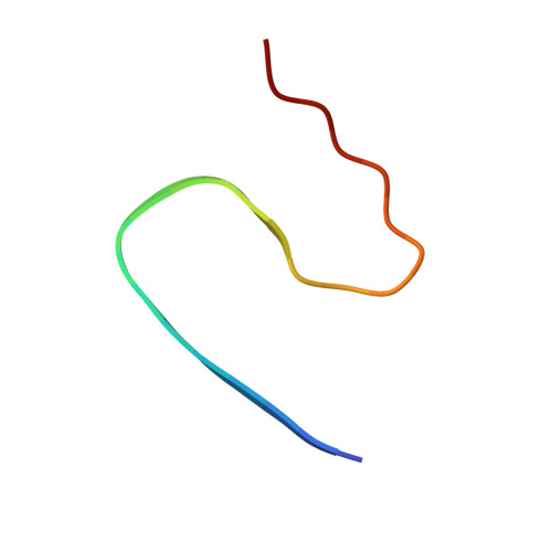
    8IB0

  • PubMed Abstract: 

    Receptor-interacting protein kinase 1 (RIPK1) is a therapeutic target in treating neurodegenerative diseases and cancers. RIPK1 has three distinct functional domains, with the center domain containing a receptor-interacting protein homotypic interaction motif (RHIM), which mediates amyloid formation. The functional amyloid formed by RIPK1 and/or RIPK3 is a crucial intermediate in regulating cell necroptosis. In this study, the amyloid structure of mouse RIPK1, formed by an 82-residue sequence centered at RHIM, is presented. It reveals the "N"-shaped folding of the protein subunit in the fibril with four β-strands. The folding pattern is shared by several amyloid structures formed by proteins with RHIM, with the central β-strand formed by the most conserved tetrad sequence I/VQI/VG. However, the solid-state NMR results indicate a structural difference between mouse RIPK1 and mouse RIPK3. A change in the structural rigidity is also suggested by the observation of weakened signals for mouse RIPK3 upon mixing with RIPK1 to form the RIPK1/RIPK3 complex fibrils. Our results provide vital information to understand the interactions between different proteins with RHIM, which will help us further comprehend the regulation mechanism in cell necroptosis.


  • Organizational Affiliation
    • School of Life Science and Technology, ShanghaiTech University, Shanghai, 201210, China.

Macromolecule Content 

  • Total Structure Weight: 13.51 kDa 
  • Atom Count: 950 
  • Modeled Residue Count: 120 
  • Deposited Residue Count: 120 
  • Unique protein chains: 1

Macromolecules

Find similar proteins by:  Sequence   |   3D Structure  
Entity ID: 1
MoleculeChains  Sequence LengthOrganismDetailsImage
Receptor-interacting serine/threonine-protein kinase 1
A, B, C, D, E
24Mus musculusMutation(s): 0 
Gene Names: Ripk1RinpRip
EC: 2.7.11.1
UniProt
Find proteins for Q60855 (Mus musculus)
Explore Q60855 
Go to UniProtKB:  Q60855
Entity Groups
Sequence Clusters30% Identity50% Identity70% Identity90% Identity95% Identity100% Identity
UniProt GroupQ60855
Sequence Annotations
Expand
Reference Sequence

Experimental Data & Validation

Experimental Data

  • Method: SOLID-STATE NMR
  • Conformers Calculated: 196 
  • Conformers Submitted: 10 
  • Selection Criteria: structures with the lowest energy 

Structure Validation

View Full Validation Report


Entry History 

& Funding Information

Deposition Data


Funding OrganizationLocationGrant Number
National Natural Science Foundation of China (NSFC)China32171185

Revision History  (Full details and data files)

  • Version 1.0: 2023-03-22
    Type: Initial release
  • Version 1.1: 2024-05-15
    Changes: Data collection, Database references
  • Version 1.2: 2024-08-21
    Changes: Database references
  • Version 1.3: 2024-08-28
    Changes: Database references