


Analysis of the catalytic mechanism of meso-DAPDH and extension of D-aromatic amino acid substrate scope
Wu, T.F., Song, W.To be published.
Experimental Data Snapshot
Starting Model: experimental
View more details
Entity ID: 1 | |||||
|---|---|---|---|---|---|
| Molecule | Chains | Sequence Length | Organism | Details | Image |
| Meso-diaminopimelate D-dehydrogenase | 299 | Proteus vulgaris | Mutation(s): 0 Gene Names: EKQ45_05540 EC: 1.4.1.16 | 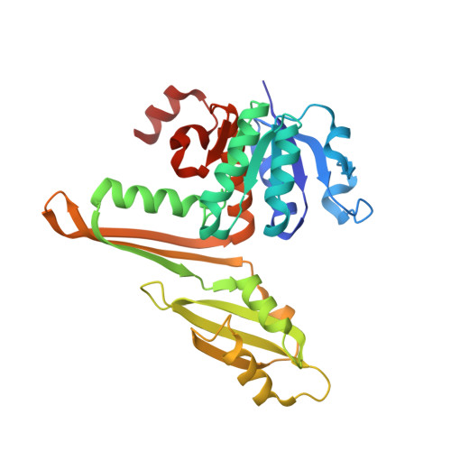 | |
UniProt | |||||
Entity Groups | |||||
| Sequence Clusters | 30% Identity50% Identity70% Identity90% Identity95% Identity100% Identity | ||||
| UniProt Group | A0A379F6K8 | ||||
Sequence AnnotationsExpand | |||||
Reference Sequence | |||||
| Ligands 2 Unique | |||||
|---|---|---|---|---|---|
| ID | Chains | Name / Formula / InChI Key | 2D Diagram | 3D Interactions | |
| API (Subject of Investigation/LOI) Download:Ideal Coordinates CCD File | E [auth A] F [auth A] K [auth B] L [auth B] P [auth C] | 2,6-DIAMINOPIMELIC ACID C7 H14 N2 O4 GMKMEZVLHJARHF-SYDPRGILSA-N |  | ||
| SO4 Download:Ideal Coordinates CCD File | AA [auth D] G [auth A] H [auth A] I [auth A] J [auth A] | SULFATE ION O4 S QAOWNCQODCNURD-UHFFFAOYSA-L |  | ||
| Length ( Å ) | Angle ( ˚ ) |
|---|---|
| a = 213.3 | α = 90 |
| b = 213.3 | β = 90 |
| c = 245.84 | γ = 120 |
| Software Name | Purpose |
|---|---|
| PHENIX | refinement |
| Funding Organization | Location | Grant Number |
|---|---|---|
| Not funded | -- |


