


Crystal structure of VIM-2 Metallo-beta-lactamase in complex with 10-HHIA
Wachino, J.To be published.
Experimental Data Snapshot
Starting Model: experimental
View more details
Entity ID: 1 | |||||
|---|---|---|---|---|---|
| Molecule | Chains | Sequence Length | Organism | Details | Image |
| Beta-lactamase class B VIM-2 | A, B [auth D] | 246 | Pseudomonas aeruginosa | Mutation(s): 0 Gene Names: blaVIM-2, bla vim-2, bla-VIM-2, blasVIM-2, blaVIM2, blm, VIM-2, PAERUG_P32_London_17_VIM_2_10_11_06255 EC: 3.5.2.6 | 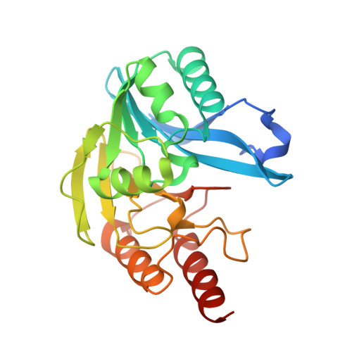 |
UniProt | |||||
Entity Groups | |||||
| Sequence Clusters | 30% Identity50% Identity70% Identity90% Identity95% Identity100% Identity | ||||
| UniProt Group | Q9K2N0 | ||||
Sequence AnnotationsExpand | |||||
Reference Sequence | |||||
| Ligands 3 Unique | |||||
|---|---|---|---|---|---|
| ID | Chains | Name / Formula / InChI Key | 2D Diagram | 3D Interactions | |
| OR6 (Subject of Investigation/LOI) Download:Ideal Coordinates CCD File | M [auth D] | (2~{S})-2-butyl-3-methylidene-butanedioic acid C9 H14 O4 ULDVOPUXMRCBDH-ZETCQYMHSA-N |  | ||
| ZN Download:Ideal Coordinates CCD File | C [auth A] D [auth A] E [auth A] H [auth D] I [auth D] | ZINC ION Zn PTFCDOFLOPIGGS-UHFFFAOYSA-N |  | ||
| FMT Download:Ideal Coordinates CCD File | F [auth A], G [auth A], K [auth D], L [auth D] | FORMIC ACID C H2 O2 BDAGIHXWWSANSR-UHFFFAOYSA-N |  | ||
| Length ( Å ) | Angle ( ˚ ) |
|---|---|
| a = 101.32 | α = 90 |
| b = 79.25 | β = 130.14 |
| c = 67.9 | γ = 90 |
| Software Name | Purpose |
|---|---|
| REFMAC | refinement |
| iMOSFLM | data reduction |
| SCALA | data scaling |
| MOLREP | phasing |
| Funding Organization | Location | Grant Number |
|---|---|---|
| Not funded | -- |


