Evaluation of the Inhibition Potency of Nirmatrelvir against Main Protease Mutants of SARS-CoV-2 Variants.
Jiang, H., Zhou, Y., Zou, X., Hu, X., Wang, J., Zeng, P., Li, W., Zeng, X., Zhang, J., Li, J.(2023) Biochemistry 62: 2055-2064
- PubMed: 37222536 Search on PubMed
- DOI: https://doi.org/10.1021/acs.biochem.3c00075
- Primary Citation Related Structures:
8HVK, 8HVL, 8HVM, 8HVN, 8HVO, 8HZR - PubMed Abstract:
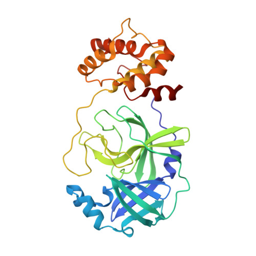
SARS-CoV-2 continues to pose a threat to public health. Main protease (M pro ) is one of the most lucrative drug targets for developing specific antivirals against SARS-CoV-2 infection. By targeting M pro , peptidomimetic nirmatrelvir is able to inhibit viral replication of SARS-CoV-2 and reduce the risk for progression to severe COVID-19. However, multiple mutations in the gene encoding M pro of emerging SARS-CoV-2 variants raise a concern of drug resistance. In the present study, we expressed 16 previously reported SARS-CoV-2 M pro mutants (G15S, T25I, T45I, S46F, S46P, D48N, M49I, L50F, L89F, K90R, P132H, N142S, V186F, R188K, T190I, and A191V). We evaluated the inhibition potency of nirmatrelvir against these M pro mutants and solved the crystal structures of representative M pro mutants of SARS-CoV-2 bound to nirmatrelvir. Enzymatic inhibition assays revealed that these M pro variants remain susceptible to nirmatrelvir as the wildtype. Detailed analysis and structural comparison provided the inhibition mechanism of M pro mutants by nirmatrelvir. These results informed the ongoing genomic surveillance of drug resistance of emerging SARS-CoV-2 variants to nirmatrelvir and facilitate the development of next-generation anticoronavirus drugs.
- School of Basic Medical Sciences, Nanchang University, Nanchang 330031, China.
Organizational Affiliation: