Crystal Structure of Glycoside Hydrolase Family 20 Lacto- N -biosidase from Soil Bacterium Streptomyces sp. Strain 142.
Fujio, N., Fushinobu, S., Yamada, C.(2025) J Appl Glycosci (1999) 72: 7202101-7202101
- PubMed: 40502377 Search on PubMedSearch on PubMed Central
- DOI: https://doi.org/10.5458/jag.7202101
- Primary Citation Related Structures:
8HVB, 8HVC, 8HVD - PubMed Abstract:
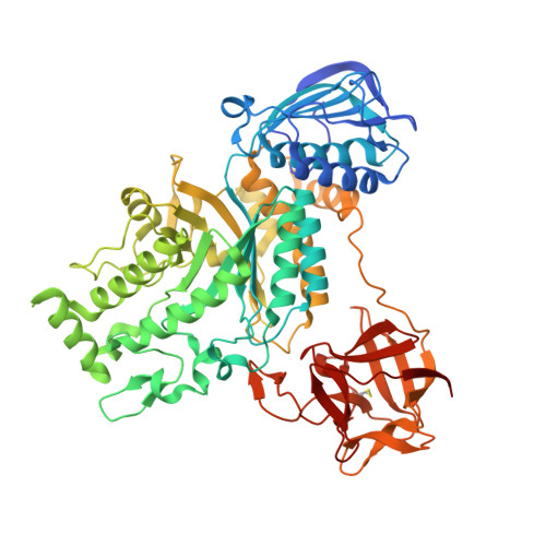
Lacto- N -biosidase hydrolyzes the β-GlcNAc or β-GalNAc bond of sugar chains to release lacto- N -biose I (Gal-β1,3-GlcNAc) or galacto- N -biose (Gal-β1,3-GalNAc) from the non-reducing end. Typical substrates for lacto- N -biosidase include type I oligosaccharides contained in human breast milk, such as lacto- N -tetraose. Lacto- N -biosidases have recently received significant attention because of their potential to synthesize milk oligosaccharides. Bifidobacterial lacto- N -biosidases belonging to glycoside hydrolase families 20 and 136 have been studied. The GH20 lacto- N -biosidases utilize a substrate-associated hydrolysis mechanism. LnbB from Bifidobacterium bifidum is the only lacto- N -biosidase with reported crystal structures in GH20. In this study, the crystal structure of the lacto- N -biosidase from Streptomyces sp. strain 142 ( Str LNBase) was solved in a complex with lacto- N -biose and galacto- N -biose. The stabilizing residue, which recognizes the nitrogen atom of the N -acetyl group of the -1 subsite, and the catalytic acid/base residue, were determined to be D304 and E305, respectively. The structure of Str LNBase is similar to that of LnbB; however, in the complex with galacto- N -biose, there were two structures exhibiting different sugar conformations. A phylogenetic analysis revealed that lacto- N -biosidases discovered in the soil bacteria Streptomyces spp. and human gut bacteria Bifidobacterium spp. may be divided into two separate groups, which suggests that they evolved divergently.
- 1 Department of Biotechnology, The University of Tokyo.
Organizational Affiliation: