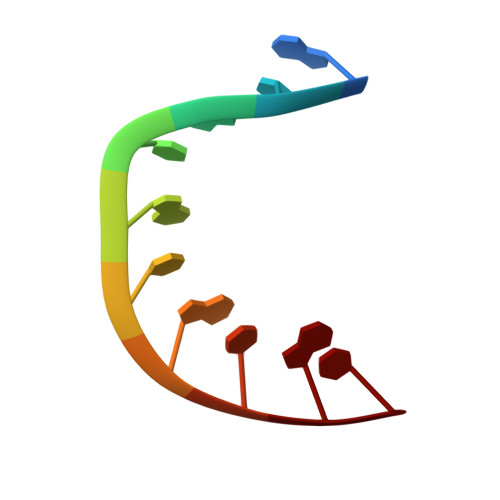
8HIS

Crystal structure of DNA decamer containing GuNA[Me,tBu]

  • Classification: DNA
  • Organism(s): synthetic construct
  • Mutation(s): No 

  • Deposited: 2022-11-21 Released: 2023-08-09 
  • Deposition Author(s): Aoyama, H., Obika, S., Yamaguchi, T.
  • Funding Organization(s): Japan Society for the Promotion of Science (JSPS), Japan Agency for Medical Research and Development (AMED)

Experimental Data Snapshot

  • Method: X-RAY DIFFRACTION
  • Resolution: 2.01 Å
  • R-Value Free: 0.228 
  • R-Value Work: 0.178 
  • R-Value Observed: 0.183 

wwPDB Validation   3D Report Full Report


This is version 1.1 of the entry. See complete history


Literature

Mechanism of the extremely high duplex-forming ability of oligonucleotides modified with N-tert-butylguanidine- or N-tert-butyl-N'- methylguanidine-bridged nucleic acids.

Yamaguchi, T.Horie, N.Aoyama, H.Kumagai, S.Obika, S.

(2023) Nucleic Acids Res 51: 7749-7761

  • DOI: https://doi.org/10.1093/nar/gkad608
  • Primary Citation of Related Structures:  
    8HIS, 8HU5, 8I50

  • PubMed Abstract: 

    Antisense oligonucleotides (ASOs) are becoming a promising class of drugs for treating various diseases. Over the past few decades, many modified nucleic acids have been developed for application to ASOs, aiming to enhance their duplex-forming ability toward cognate mRNA and improve their stability against enzymatic degradations. Modulating the sugar conformation of nucleic acids by substituting an electron-withdrawing group at the 2'-position or incorporating a 2',4'-bridging structure is a common approach for enhancing duplex-forming ability. Here, we report on incorporating an N-tert-butylguanidinium group at the 2',4'-bridging structure, which greatly enhances duplex-forming ability because of its interactions with the minor groove. Our results indicated that hydrophobic substituents fitting the grooves of duplexes also have great potential to increase duplex-forming ability.


  • Organizational Affiliation

    Graduate School of Pharmaceutical Sciences, Osaka University, 1-6 Yamadaoka, Suita, Osaka 565-0871, Japan.


Macromolecules

Find similar nucleic acids by:  Sequence   |   3D Structure  

Entity ID: 1
MoleculeChains LengthOrganismImage
DNA (5'-D(*GP*CP*GP*TP*AP*(LR6)P*AP*CP*GP*C)-3')
A, B
10synthetic construct
Sequence Annotations
Expand
  • Reference Sequence
Small Molecules
Ligands 1 Unique
IDChains Name / Formula / InChI Key2D Diagram3D Interactions
CAD
Query on CAD

Download Ideal Coordinates CCD File 
C [auth B]CACODYLIC ACID
C2 H7 As O2
OGGXGZAMXPVRFZ-UHFFFAOYSA-N
Experimental Data & Validation

Experimental Data

  • Method: X-RAY DIFFRACTION
  • Resolution: 2.01 Å
  • R-Value Free: 0.228 
  • R-Value Work: 0.178 
  • R-Value Observed: 0.183 
  • Space Group: P 21 21 21
Unit Cell:
Length ( Å )Angle ( ˚ )
a = 25.07α = 90
b = 44.36β = 90
c = 45.19γ = 90
Software Package:
Software NamePurpose
PHENIXrefinement
XDSdata reduction
Aimlessdata scaling
PHASERphasing
PDB_EXTRACTdata extraction

Structure Validation

View Full Validation Report


Entry History & Funding Information

Deposition Data


Funding OrganizationLocationGrant Number
Japan Society for the Promotion of Science (JSPS)Japan20K05748
Japan Society for the Promotion of Science (JSPS)Japan21K06511
Japan Agency for Medical Research and Development (AMED)JapanJP19am0101084
Japan Agency for Medical Research and Development (AMED)JapanJP18am0301004
Japan Agency for Medical Research and Development (AMED)Japan19am0401003
Japan Agency for Medical Research and Development (AMED)JapanJP21ae0121022
Japan Agency for Medical Research and Development (AMED)JapanJP21ae0121023
Japan Agency for Medical Research and Development (AMED)JapanJP21ae012102

Revision History  (Full details and data files)

  • Version 1.0: 2023-08-09
    Type: Initial release
  • Version 1.1: 2023-09-06
    Changes: Data collection, Database references