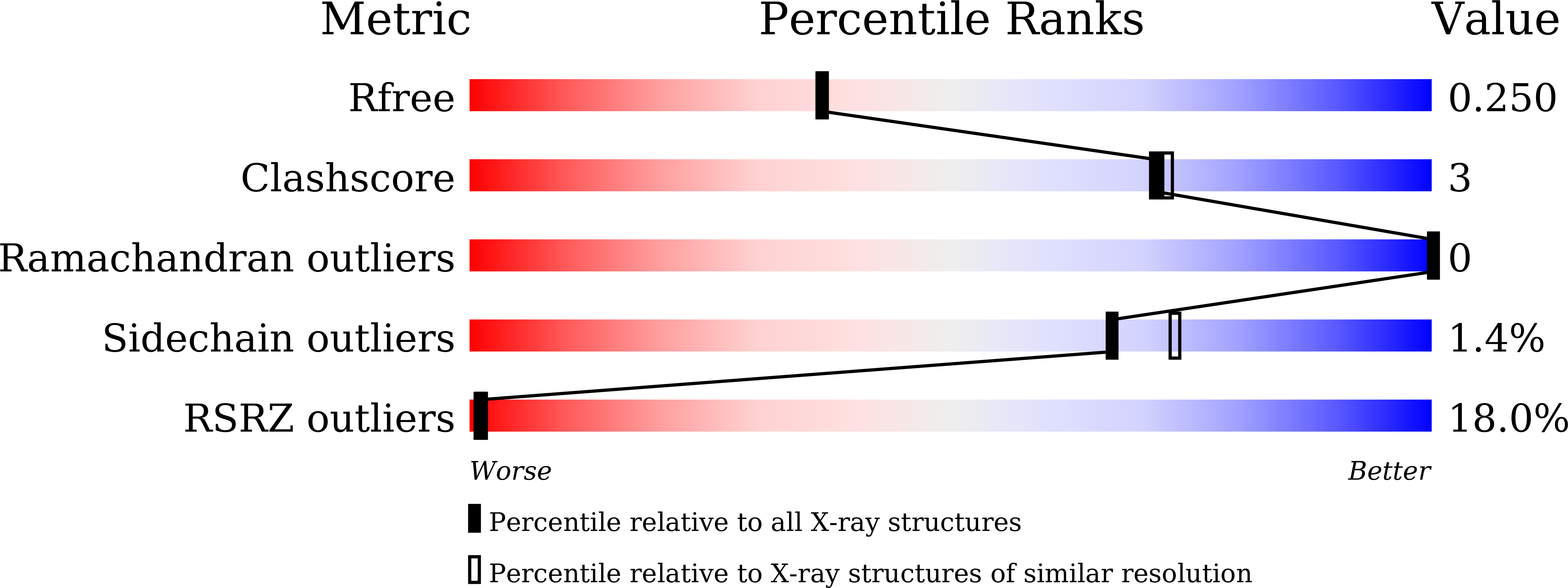
 8HGQ | pdb_00008hgq

The apo-flavodoxin dimer from Synechococcus elongatus PCC 7942


Experimental Data Snapshot

  • Method: X-RAY DIFFRACTION
  • Resolution: 2.09 Å
  • R-Value Free: 
    0.239 (Depositor), 0.250 (DCC) 
  • R-Value Work: 
    0.192 (Depositor), 0.204 (DCC) 
  • R-Value Observed: 
    0.194 (Depositor) 

Starting Model: experimental
View more details

wwPDB Validation 3D Report Full Report

Validation slider image for 8HGQ

This is version 1.2 of the entry. See complete history. 

Literature

A dimer-monomer transition captured by the crystal structures of cyanobacterial apo flavodoxin.

Liu, S., Chen, Y., Du, T., Zhao, W., Liu, X., Zhang, H., Yuan, Q., Gao, L., Dong, Y., Gao, X., Gong, Y., Cao, P.

(2022) Biochem Biophys Res Commun 639: 134-141

  • DOI: https://doi.org/10.1016/j.bbrc.2022.11.089
  • Primary Citation Related Structures: 
    8HGQ, 8HGR

  • PubMed Abstract: 

    In cyanobacteria and algae (but not plants), flavodoxin (Fld) replaces ferredoxin (Fd) under stress conditions to transfer electrons from photosystem I (PSI) to ferredoxin-NADP + reductase (FNR) during photosynthesis. Fld constitutes a small electron carrier noncovalently bound to flavin mononucleotide (FMN), and also an ideal model for revealing the protein/flavin-binding mechanism because of its relative simplicity compared to other flavoproteins. Here, we report two crystal structures of apo-Fld from Synechococcus sp. PCC 7942, one dimeric structure of 2.09 Å and one monomeric structure of 1.84 Å resolution. Analytical ultracentrifugation showed that in solution, apo-Fld exists both as monomers and dimers. Our dimer structure contains two ligand-binding pockets separated by a distance of 45 Å, much longer than the previous structures of FMN-bound dimers. These results suggested a potential dimer-monomer transition mechanism of cyanobacterial apo-Fld. We further propose that the dimer represents the "standby" state to stabilize itself, while the monomer constitutes the "ready" state to bind FMN. Furthermore, we generated a new docking model of cyanobacterial Fld-FNR complex based on the recently reported cryo-EM structures, and mapped the special interactions between Fld and FNR in detail.


  • Organizational Affiliation: 
    • Faculty of Environment and Life, Beijing University of Technology, 100 Pingleyuan, Chaoyang District, Beijing, 100124, China.

Macromolecule Content 

  • Total Structure Weight: 20.88 kDa 
  • Atom Count: 1,439 
  • Modeled Residue Count: 172 
  • Deposited Residue Count: 186 
  • Unique protein chains: 1

Macromolecules

Find similar proteins by:|  3D Structure
Entity ID: 1
MoleculeChains  Sequence LengthOrganismDetailsImage
Flavodoxin186Synechococcus elongatus PCC 7942 = FACHB-805Mutation(s): 0 
Gene Names: isiB, Synpcc7942_1541
UniProt
Find proteins for P10340 (Synechococcus elongatus (strain ATCC 33912 / PCC 7942 / FACHB-805))
Explore P10340 
Go to UniProtKB:  P10340
Entity Groups
Sequence Clusters30% Identity50% Identity70% Identity90% Identity95% Identity100% Identity
UniProt GroupP10340
Sequence Annotations
Expand
Reference Sequence

Small Molecules

Ligands 1 Unique
IDChains Name / Formula / InChI Key2D Diagram3D Interactions
PO4

Query on PO4


Download:Ideal Coordinates CCD File
B [auth A]PHOSPHATE ION
O4 P
NBIIXXVUZAFLBC-UHFFFAOYSA-K

Experimental Data & Validation

Experimental Data

  • Method: X-RAY DIFFRACTION
  • Resolution: 2.09 Å
  • R-Value Free:  0.239 (Depositor), 0.250 (DCC) 
  • R-Value Work:  0.192 (Depositor), 0.204 (DCC) 
  • R-Value Observed: 0.194 (Depositor) 
Space Group: P 41 21 2
Unit Cell:
Length ( Å )Angle ( ˚ )
a = 49.56α = 90
b = 49.56β = 90
c = 161.025γ = 90
Software Package:
Software NamePurpose
REFMACrefinement
PHENIXrefinement
XDSdata reduction
XDSdata scaling
PHASERphasing

Structure Validation

View Full Validation Report


Entry History 

& Funding Information

Deposition Data


Funding OrganizationLocationGrant Number
National Natural Science Foundation of China (NSFC)China32070259

Revision History  (Full details and data files)

  • Version 1.0: 2022-12-14
    Type: Initial release
  • Version 1.1: 2022-12-28
    Changes: Database references
  • Version 1.2: 2023-11-29
    Changes: Data collection, Refinement description
