8HDT

Cyanophage Pam3 capsid asymmetric unit


Experimental Data Snapshot

  • Method: ELECTRON MICROSCOPY
  • Resolution: 3.17 Å
  • Aggregation State: PARTICLE 
  • Reconstruction Method: SINGLE PARTICLE 

wwPDB Validation   3D Report Full Report


This is version 1.1 of the entry. See complete history


Literature

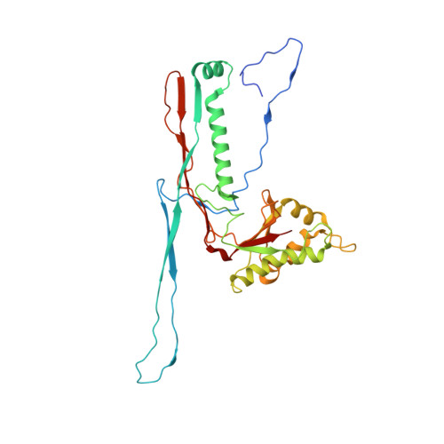
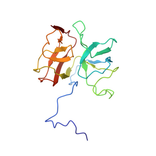
Fine structure and assembly pattern of a minimal myophage Pam3.

Yang, F.Jiang, Y.L.Zhang, J.T.Zhu, J.Du, K.Yu, R.C.Wei, Z.L.Kong, W.W.Cui, N.Li, W.F.Chen, Y.Li, Q.Zhou, C.Z.

(2023) Proc Natl Acad Sci U S A 120: e2213727120-e2213727120

  • DOI: https://doi.org/10.1073/pnas.2213727120
  • Primary Citation of Related Structures:  
    7YFW, 7YFZ, 8HDR, 8HDS, 8HDT, 8HDW

  • PubMed Abstract: 

    The myophage possesses a contractile tail that penetrates its host cell envelope. Except for investigations on the bacteriophage T4 with a rather complicated structure, the assembly pattern and tail contraction mechanism of myophage remain largely unknown. Here, we present the fine structure of a freshwater Myoviridae cyanophage Pam3, which has an icosahedral capsid of ~680 Å in diameter, connected via a three-section neck to an 840-Å-long contractile tail, ending with a three-module baseplate composed of only six protein components. This simplified baseplate consists of a central hub-spike surrounded by six wedge heterotriplexes, to which twelve tail fibers are covalently attached via disulfide bonds in alternating upward and downward configurations. In vitro reduction assays revealed a putative redox-dependent mechanism of baseplate assembly and tail sheath contraction. These findings establish a minimal myophage that might become a user-friendly chassis phage in synthetic biology.


  • Organizational Affiliation

    School of Life Sciences, Division of Life Sciences and Medicine, University of Science and Technology of China, Hefei 230026, China.


Macromolecules
Find similar proteins by:  (by identity cutoff)  |  3D Structure
Entity ID: 1
MoleculeChains Sequence LengthOrganismDetailsImage
Major capsid
A, B, C, D, E
A, B, C, D, E, F, N
310uncultured cyanophageMutation(s): 0 
UniProt
Find proteins for A0A9E7DT27 (Pseudanabaena phage Pam3)
Explore A0A9E7DT27 
Go to UniProtKB:  A0A9E7DT27
Entity Groups  
Sequence Clusters30% Identity50% Identity70% Identity90% Identity95% Identity100% Identity
UniProt GroupA0A9E7DT27
Sequence Annotations
Expand
  • Reference Sequence
Find similar proteins by:  (by identity cutoff)  |  3D Structure
Entity ID: 2
MoleculeChains Sequence LengthOrganismDetailsImage
Cement
G, H, I, J, K
G, H, I, J, K, L, M
166uncultured cyanophageMutation(s): 0 
UniProt
Find proteins for A0A9E7DT54 (Pseudanabaena phage Pam3)
Explore A0A9E7DT54 
Go to UniProtKB:  A0A9E7DT54
Entity Groups  
Sequence Clusters30% Identity50% Identity70% Identity90% Identity95% Identity100% Identity
UniProt GroupA0A9E7DT54
Sequence Annotations
Expand
  • Reference Sequence
Experimental Data & Validation

Experimental Data

  • Method: ELECTRON MICROSCOPY
  • Resolution: 3.17 Å
  • Aggregation State: PARTICLE 
  • Reconstruction Method: SINGLE PARTICLE 
EM Software:
TaskSoftware PackageVersion
RECONSTRUCTIONRELION3.1

Structure Validation

View Full Validation Report


Entry History & Funding Information

Deposition Data


Funding OrganizationLocationGrant Number
Ministry of Science and Technology (MoST, China)China2018YFA0903100

Revision History  (Full details and data files)

  • Version 1.0: 2023-01-18
    Type: Initial release
  • Version 1.1: 2023-02-01
    Changes: Database references