8HDD

Complex structure of catalytic, small, and a partial electron transfer subunits from Burkholderia cepacia FAD glucose dehydrogenase


Experimental Data Snapshot

  • Method: X-RAY DIFFRACTION
  • Resolution: 3.00 Å
  • R-Value Free: 0.323 
  • R-Value Work: 0.275 
  • R-Value Observed: 0.278 

wwPDB Validation   3D Report Full Report


Ligand Structure Quality Assessment 


This is version 1.2 of the entry. See complete history


Literature

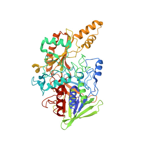
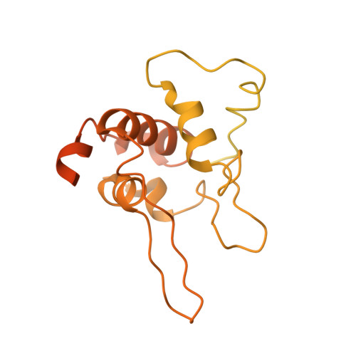
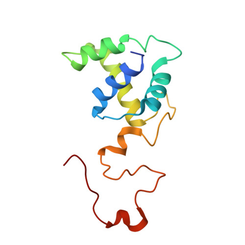
Microgravity environment grown crystal structure information based engineering of direct electron transfer type glucose dehydrogenase.

Okuda-Shimazaki, J.Yoshida, H.Lee, I.Kojima, K.Suzuki, N.Tsugawa, W.Yamada, M.Inaka, K.Tanaka, H.Sode, K.

(2022) Commun Biol 5: 1334-1334

  • DOI: https://doi.org/10.1038/s42003-022-04286-9
  • Primary Citation of Related Structures:  
    8HDD

  • PubMed Abstract: 

    The heterotrimeric flavin adenine dinucleotide dependent glucose dehydrogenase is a promising enzyme for direct electron transfer (DET) principle-based glucose sensors within continuous glucose monitoring systems. We elucidate the structure of the subunit interface of this enzyme by preparing heterotrimer complex protein crystals grown under a space microgravity environment. Based on the proposed structure, we introduce inter-subunit disulfide bonds between the small and electron transfer subunits (5 pairs), as well as the catalytic and the electron transfer subunits (9 pairs). Without compromising the enzyme's catalytic efficiency, a mutant enzyme harboring Pro205Cys in the catalytic subunit, Asp383Cys and Tyr349Cys in the electron transfer subunit, and Lys155Cys in the small subunit, is determined to be the most stable of the variants. The developed engineered enzyme demonstrate a higher catalytic activity and DET ability than the wild type. This mutant retains its full activity below 70 °C as well as after incubation at 75 °C for 15 min - much higher temperatures than the current gold standard enzyme, glucose oxidase, is capable of withstanding.


  • Organizational Affiliation

    Joint Department of Biomedical Engineering, The University of North Carolina at Chapel Hill and North Carolina State University, Chapel Hill, NC27599, USA.


Macromolecules
Find similar proteins by:  (by identity cutoff)  |  3D Structure
Entity ID: 1
MoleculeChains Sequence LengthOrganismDetailsImage
Glucose dehydrogenase539Burkholderia cepaciaMutation(s): 0 
Gene Names: gdhAlpha
EC: 1.1.5.9
UniProt
Find proteins for Q8GQE7 (Burkholderia cepacia)
Explore Q8GQE7 
Go to UniProtKB:  Q8GQE7
Entity Groups  
Sequence Clusters30% Identity50% Identity70% Identity90% Identity95% Identity100% Identity
UniProt GroupQ8GQE7
Sequence Annotations
Expand
  • Reference Sequence
Find similar proteins by:  (by identity cutoff)  |  3D Structure
Entity ID: 2
MoleculeChains Sequence LengthOrganismDetailsImage
Glucose dehydrogenase beta subunit482Burkholderia cepaciaMutation(s): 2 
UniProt
Find proteins for Q71JE9 (Burkholderia cepacia)
Explore Q71JE9 
Go to UniProtKB:  Q71JE9
Entity Groups  
Sequence Clusters30% Identity50% Identity70% Identity90% Identity95% Identity100% Identity
UniProt GroupQ71JE9
Sequence Annotations
Expand
  • Reference Sequence
Find similar proteins by:  (by identity cutoff)  |  3D Structure
Entity ID: 3
MoleculeChains Sequence LengthOrganismDetailsImage
Twin-arginine translocation pathway signal121Burkholderia cepaciaMutation(s): 0 
Gene Names: BMULJ_04411
UniProt
Find proteins for A0A0H3KLY3 (Burkholderia multivorans (strain ATCC 17616 / 249))
Explore A0A0H3KLY3 
Go to UniProtKB:  A0A0H3KLY3
Entity Groups  
Sequence Clusters30% Identity50% Identity70% Identity90% Identity95% Identity100% Identity
UniProt GroupA0A0H3KLY3
Sequence Annotations
Expand
  • Reference Sequence
Experimental Data & Validation

Experimental Data

  • Method: X-RAY DIFFRACTION
  • Resolution: 3.00 Å
  • R-Value Free: 0.323 
  • R-Value Work: 0.275 
  • R-Value Observed: 0.278 
  • Space Group: P 21 21 2
Unit Cell:
Length ( Å )Angle ( ˚ )
a = 204.015α = 90
b = 71.797β = 90
c = 114.221γ = 90
Software Package:
Software NamePurpose
XSCALEdata scaling
XDSdata reduction
REFMACrefinement
MOLREPphasing

Structure Validation

View Full Validation Report


Ligand Structure Quality Assessment 


Entry History & Funding Information

Deposition Data


Funding OrganizationLocationGrant Number
Other privateJapanNA (sponsorship research)

Revision History  (Full details and data files)

  • Version 1.0: 2022-12-14
    Type: Initial release
  • Version 1.1: 2023-11-29
    Changes: Data collection, Refinement description
  • Version 1.2: 2024-05-08
    Changes: Database references