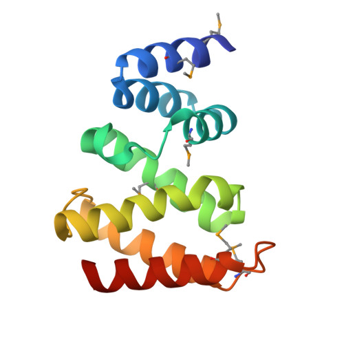
8HBR

The C-terminal domain of Spiral2


Experimental Data Snapshot

  • Method: X-RAY DIFFRACTION
  • Resolution: 2.20 Å
  • R-Value Free: 0.256 
  • R-Value Work: 0.202 
  • R-Value Observed: 0.205 

wwPDB Validation   3D Report Full Report


This is version 1.1 of the entry. See complete history


Literature

Crystal structure of the C-terminal domain of the plant-specific microtubule-associated protein Spiral2.

Ohno, M.Higuchi, Y.Hayashi, I.

(2023) Acta Crystallogr F Struct Biol Commun 79: 17-22

  • DOI: https://doi.org/10.1107/S2053230X22011815
  • Primary Citation of Related Structures:  
    8HBR

  • PubMed Abstract: 

    Plant cells form microtubule arrays, called `cortical microtubules', beneath the plasma membrane which are critical for cell-wall organization and directional cell growth. Cortical microtubules are nucleated independently of centrosomes. Spiral2 is a land-plant-specific microtubule minus-end-targeting protein that stabilizes the minus ends by inhibiting depolymerization of the filament. Spiral2 possesses an N-terminal microtubule-binding domain and a conserved C-terminal domain whose function is unknown. In this study, the crystal structure of the conserved C-terminal domain of Spiral2 was determined using the single-wavelength anomalous dispersion method. Refinement of the model to a resolution of 2.2 Å revealed a helix-turn-helix fold with seven α-helices. The protein crystallized as a dimer, but SEC-MALS analysis showed the protein to be monomeric. A structural homology search revealed that the protein has similarity to the C-terminal domain of the katanin regulatory subunit p80. The structure presented here suggests that the C-terminal domain of Spiral2 represents a new class of microtubule dynamics modulator across the kingdom.


  • Organizational Affiliation

    Department of Medical Life Science, Yokohama City University, 1-7-29 Suehiro, Tsurumi, Yokohama, Kanagawa 230-0045, Japan.


Macromolecules
Find similar proteins by:  (by identity cutoff)  |  3D Structure
Entity ID: 1
MoleculeChains Sequence LengthOrganismDetailsImage
TOG domain-containing protein
A, B, C, D, E
A, B, C, D, E, F
135Physcomitrium patensMutation(s): 0 
Gene Names: PHYPA_024812
UniProt
Find proteins for A0A7I4C2S7 (Physcomitrium patens)
Explore A0A7I4C2S7 
Go to UniProtKB:  A0A7I4C2S7
Entity Groups  
Sequence Clusters30% Identity50% Identity70% Identity90% Identity95% Identity100% Identity
UniProt GroupA0A7I4C2S7
Sequence Annotations
Expand
  • Reference Sequence
Small Molecules
Modified Residues  1 Unique
IDChains TypeFormula2D DiagramParent
MSE
Query on MSE
A, B, C, D, E
A, B, C, D, E, F
L-PEPTIDE LINKINGC5 H11 N O2 SeMET
Experimental Data & Validation

Experimental Data

  • Method: X-RAY DIFFRACTION
  • Resolution: 2.20 Å
  • R-Value Free: 0.256 
  • R-Value Work: 0.202 
  • R-Value Observed: 0.205 
  • Space Group: P 1 21 1
Unit Cell:
Length ( Å )Angle ( ˚ )
a = 55.509α = 90
b = 109.56β = 113.2
c = 69.787γ = 90
Software Package:
Software NamePurpose
REFMACrefinement
XDSdata reduction
pointlessdata scaling
CRANK2phasing

Structure Validation

View Full Validation Report


Entry History & Funding Information

Deposition Data

  • Released Date: 2023-01-18 
  • Deposition Author(s): Hayashi, I.

Funding OrganizationLocationGrant Number
Japan Society for the Promotion of Science (JSPS)Japan--

Revision History  (Full details and data files)

  • Version 1.0: 2023-01-18
    Type: Initial release
  • Version 1.1: 2024-10-09
    Changes: Data collection, Structure summary