8HB5 | pdb_00008hb5

Crystal structure of Mincle in complex with HD-275


Experimental Data Snapshot

  • Method: X-RAY DIFFRACTION
  • Resolution: 2.60 Å
  • R-Value Free: 
    0.336 (Depositor), 0.335 (DCC) 
  • R-Value Work: 
    0.296 (Depositor), 0.296 (DCC) 
  • R-Value Observed: 
    0.298 (Depositor) 

Starting Model: experimental
View more details

wwPDB Validation 3D Report Full Report

Validation slider image for 8HB5

Ligand Structure Quality Assessment 


This is version 1.2 of the entry. See complete history

Literature

PGL-III, a Rare Intermediate of Mycobacterium leprae Phenolic Glycolipid Biosynthesis, Is a Potent Mincle Ligand.

Ishizuka, S.van Dijk, J.H.M.Kawakita, T.Miyamoto, Y.Maeda, Y.Goto, M.Le Calvez, G.Groot, L.M.Witte, M.D.Minnaard, A.J.van der Marel, G.A.Ato, M.Nagae, M.Codee, J.D.C.Yamasaki, S.

(2023) ACS Cent Sci 9: 1388-1399

  • DOI: https://doi.org/10.1021/acscentsci.3c00040
  • Primary Citation Related Structures: 
    8H4V, 8HB5

  • PubMed Abstract: 

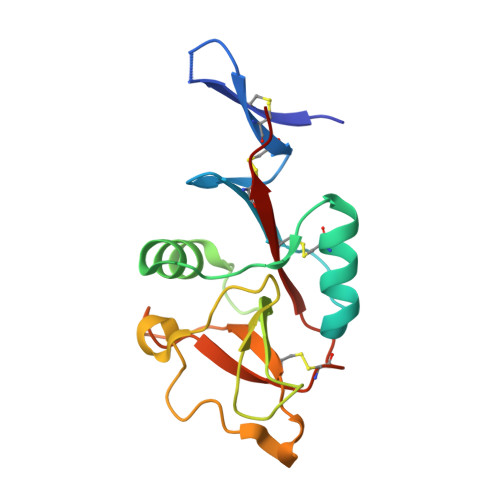
    Although leprosy (Hansen's disease) is one of the oldest known diseases, the pathogenicity of Mycobacterium leprae ( M. leprae ) remains enigmatic. Indeed, the cell wall components responsible for the immune response against M. leprae are as yet largely unidentified. We reveal here phenolic glycolipid-III (PGL-III) as an M. leprae -specific ligand for the immune receptor Mincle. PGL-III is a scarcely present trisaccharide intermediate in the biosynthetic pathway to PGL-I, an abundant and characteristic M. leprae glycolipid. Using activity-based purification, we identified PGL-III as a Mincle ligand that is more potent than the well-known M. tuberculosis trehalose dimycolate. The cocrystal structure of Mincle and a synthetic PGL-III analogue revealed a unique recognition mode, implying that it can engage multiple Mincle molecules. In Mincle-deficient mice infected with M. leprae , increased bacterial burden with gross pathologies were observed. These results show that PGL-III is a noncanonical ligand recognized by Mincle, triggering protective immunity.


  • Organizational Affiliation
    • Department of Molecular Immunology, Research Institute for Microbial Diseases, Osaka University, 3-1 Yamadaoka, Suita, Osaka 565-0871, Japan.

Macromolecule Content 

  • Total Structure Weight: 17.76 kDa 
  • Atom Count: 1,213 
  • Modeled Residue Count: 146 
  • Deposited Residue Count: 151 
  • Unique protein chains: 1

Macromolecules

Find similar proteins by:|  3D Structure
Entity ID: 1
MoleculeChains  Sequence LengthOrganismDetailsImage
C-type lectin domain family 4 member E151Bos taurusMutation(s): 1 
Gene Names: CLEC4E
UniProt
Find proteins for E1BHM0 (Bos taurus)
Explore E1BHM0 
Go to UniProtKB:  E1BHM0
Entity Groups
Sequence Clusters30% Identity50% Identity70% Identity90% Identity95% Identity100% Identity
UniProt GroupE1BHM0
Sequence Annotations
Expand
Reference Sequence

Experimental Data & Validation

Experimental Data

  • Method: X-RAY DIFFRACTION
  • Resolution: 2.60 Å
  • R-Value Free:  0.336 (Depositor), 0.335 (DCC) 
  • R-Value Work:  0.296 (Depositor), 0.296 (DCC) 
  • R-Value Observed: 0.298 (Depositor) 
Space Group: P 31 2 1
Unit Cell:
Length ( Å )Angle ( ˚ )
a = 97.515α = 90
b = 97.515β = 90
c = 46.49γ = 120
Software Package:
Software NamePurpose
PHENIXrefinement
XDSdata reduction
Aimlessdata scaling
MOLREPphasing

Structure Validation

View Full Validation Report


Ligand Structure Quality Assessment 


Entry History 

& Funding Information

Deposition Data


Funding OrganizationLocationGrant Number
Japan Society for the Promotion of Science (JSPS)Japan20H00505
Japan Society for the Promotion of Science (JSPS)Japan20K06575

Revision History  (Full details and data files)

  • Version 1.0: 2023-08-09
    Type: Initial release
  • Version 1.1: 2024-11-06
    Changes: Data collection, Structure summary
  • Version 1.2: 2026-03-04
    Changes: Refinement description