8HAE | pdb_00008hae

Cryo-EM structure of HACE1 dimer


Experimental Data Snapshot

  • Method: ELECTRON MICROSCOPY
  • Resolution: 4.55 Å
  • Aggregation State: PARTICLE 
  • Reconstruction Method: SINGLE PARTICLE 

wwPDB Validation 3D Report Full Report

Validation slider image for 8HAE

This is version 1.3 of the entry. See complete history

Literature

Structural Basis for the Enzymatic Activity of the HACE1 HECT-Type E3 Ligase Through N-Terminal Helix Dimerization.

Singh, S.Machida, S.Tulsian, N.K.Choong, Y.K.Ng, J.Shankar, S.Liu, Y.Chandiramani, K.V.Shi, J.Sivaraman, J.

(2023) Adv Sci (Weinh) 10: e2207672-e2207672

  • DOI: https://doi.org/10.1002/advs.202207672
  • Primary Citation Related Structures: 
    8H8X, 8HAE

  • PubMed Abstract: 

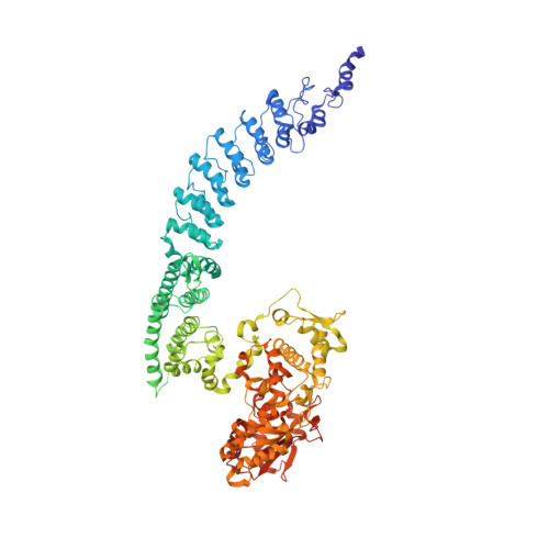
    HACE1 is an ankyrin repeat (AKR) containing HECT-type E3 ubiquitin ligase that interacts with and ubiquitinates multiple substrates. While HACE1 is a well-known tumor suppressor, its structure and mode of ubiquitination are not understood. The authors present the cryo-EM structures of human HACE1 along with in vitro functional studies that provide insights into how the enzymatic activity of HACE1 is regulated. HACE1 comprises of an N-terminal AKR domain, a middle (MID) domain, and a C-terminal HECT domain. Its unique G-shaped architecture interacts as a homodimer, with monomers arranged in an antiparallel manner. In this dimeric arrangement, HACE1 ubiquitination activity is hampered, as the N-terminal helix of one monomer restricts access to the C-terminal domain of the other. The in vitro ubiquitination assays, hydrogen-deuterium exchange mass spectrometry (HDX-MS) analysis, mutagenesis, and in silico modeling suggest that the HACE1 MID domain plays a crucial role along with the AKRs in RAC1 substrate recognition.


  • Organizational Affiliation
    • Department of Biological Sciences, National University of Singapore, 14 Science Drive 4, Singapore, 117558, Singapore.

Macromolecule Content 

  • Total Structure Weight: 204.9 kDa 
  • Atom Count: 13,426 
  • Modeled Residue Count: 1,690 
  • Deposited Residue Count: 1,818 
  • Unique protein chains: 1

Macromolecules

Find similar proteins by:|  3D Structure
Entity ID: 1
MoleculeChains  Sequence LengthOrganismDetailsImage
E3 ubiquitin-protein ligase HACE1
A, B
909Homo sapiensMutation(s): 0 
Gene Names: HACE1KIAA1320
EC: 2.3.2.26
UniProt & NIH Common Fund Data Resources
Find proteins for Q8IYU2 (Homo sapiens)
Explore Q8IYU2 
Go to UniProtKB:  Q8IYU2
PHAROS:  Q8IYU2
GTEx:  ENSG00000085382 
Entity Groups
Sequence Clusters30% Identity50% Identity70% Identity90% Identity95% Identity100% Identity
UniProt GroupQ8IYU2
Sequence Annotations
Expand
Reference Sequence

Experimental Data & Validation

Experimental Data

  • Method: ELECTRON MICROSCOPY
  • Resolution: 4.55 Å
  • Aggregation State: PARTICLE 
  • Reconstruction Method: SINGLE PARTICLE 
EM Software:
TaskSoftware PackageVersion
MODEL REFINEMENTPHENIX1.13_2998:

Structure Validation

View Full Validation Report


Entry History 

& Funding Information

Deposition Data


Funding OrganizationLocationGrant Number
Ministry of Education (MoE, Singapore)Singapore--

Revision History  (Full details and data files)

  • Version 1.0: 2023-06-28
    Type: Initial release
  • Version 1.1: 2023-08-16
    Changes: Data collection, Database references
  • Version 1.2: 2023-08-30
    Changes: Database references
  • Version 1.3: 2023-10-04
    Changes: Database references