8H3T | pdb_00008h3t

The crystal structure of AlpH


Experimental Data Snapshot

  • Method: X-RAY DIFFRACTION
  • Resolution: 1.87 Å
  • R-Value Free: 
    0.201 (Depositor), 0.201 (DCC) 
  • R-Value Work: 
    0.162 (Depositor), 0.164 (DCC) 
  • R-Value Observed: 
    0.164 (Depositor) 

Starting Model: other
View more details

wwPDB Validation 3D Report Full Report

Validation slider image for 8H3T

Ligand Structure Quality Assessment 


This is version 1.2 of the entry. See complete history

Literature

O-methyltransferase-like enzyme catalyzed diazo installation in polyketide biosynthesis.

Zhao, Y.Liu, X.Xiao, Z.Zhou, J.Song, X.Wang, X.Hu, L.Wang, Y.Sun, P.Wang, W.He, X.Lin, S.Deng, Z.Pan, L.Jiang, M.

(2023) Nat Commun 14: 5372-5372

  • DOI: https://doi.org/10.1038/s41467-023-41062-7
  • Primary Citation Related Structures: 
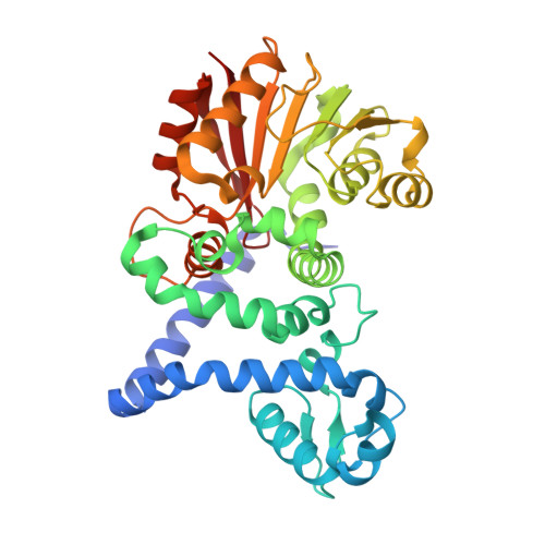
    8H3T

  • PubMed Abstract: 

    Diazo compounds are rare natural products possessing various biological activities. Kinamycin and lomaiviticin, two diazo natural products featured by the diazobenzofluorene core, exhibit exceptional potency as chemotherapeutic agents. Despite the extensive studies on their biosynthetic gene clusters and the assembly of their polyketide scaffolds, the formation of the characteristic diazo group remains elusive. L-Glutamylhydrazine was recently shown to be the hydrazine donor in kinamycin biosynthesis, however, the mechanism for the installation of the hydrazine group onto the kinamycin scaffold is still unclear. Here we describe an O-methyltransferase-like protein, AlpH, which is responsible for the hydrazine incorporation in kinamycin biosynthesis. AlpH catalyses a unique SAM-independent coupling of L-glutamylhydrazine and polyketide intermediate via a rare Mannich reaction in polyketide biosynthesis. Our discovery expands the catalytic diversity of O-methyltransferase-like enzymes and lays a strong foundation for the discovery and development of novel diazo natural products through genome mining and synthetic biology.


  • Organizational Affiliation
    • State Key Laboratory of Microbial Metabolism, Joint International Research Laboratory of Metabolic & Developmental Sciences, and School of Life Sciences and Biotechnology, Shanghai Jiao Tong University, 200030, Shanghai, P. R. China.

Macromolecule Content 

  • Total Structure Weight: 77.37 kDa 
  • Atom Count: 6,185 
  • Modeled Residue Count: 718 
  • Deposited Residue Count: 722 
  • Unique protein chains: 1

Macromolecules

Find similar proteins by:|  3D Structure
Entity ID: 1
MoleculeChains  Sequence LengthOrganismDetailsImage
AlpH
A, B
361Streptomyces galtieriMutation(s): 0 

Small Molecules

Ligands 1 Unique
IDChains Name / Formula / InChI Key2D Diagram3D Interactions
GOL
(Subject of Investigation/LOI)

Query on GOL


Download:Ideal Coordinates CCD File
C [auth B]GLYCEROL
C3 H8 O3
PEDCQBHIVMGVHV-UHFFFAOYSA-N

Experimental Data & Validation

Experimental Data

  • Method: X-RAY DIFFRACTION
  • Resolution: 1.87 Å
  • R-Value Free:  0.201 (Depositor), 0.201 (DCC) 
  • R-Value Work:  0.162 (Depositor), 0.164 (DCC) 
  • R-Value Observed: 0.164 (Depositor) 
Space Group: P 21 21 21
Unit Cell:
Length ( Å )Angle ( ˚ )
a = 68.209α = 90
b = 94.812β = 90
c = 110.108γ = 90
Software Package:
Software NamePurpose
PHENIXrefinement
PDB_EXTRACTdata extraction
autoPROCdata reduction
autoPROCdata scaling

Structure Validation

View Full Validation Report


Ligand Structure Quality Assessment 


Entry History 

& Funding Information

Deposition Data


Funding OrganizationLocationGrant Number
National Natural Science Foundation of China (NSFC)China21822705

Revision History  (Full details and data files)

  • Version 1.0: 2023-09-13
    Type: Initial release
  • Version 1.1: 2024-02-14
    Changes: Database references
  • Version 1.2: 2026-03-11
    Changes: Refinement description, Structure summary