Structural insights into the biochemical mechanism of the E2/E3 hybrid enzyme UBE2O.
Huang, H., Zhu, W., Huang, B., Fu, Z., Xiong, Y., Cao, D., Ye, Y., Chang, Q., Li, W., Li, L., Zhou, H., Niu, X., Zhang, W.(2025) Structure 33: 274-288.e4
- PubMed: 39740670 Search on PubMed
- DOI: https://doi.org/10.1016/j.str.2024.12.002
- Primary Citation Related Structures:
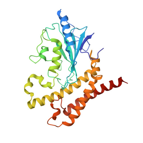
7YW1, 8GXR - PubMed Abstract:
The E2/E3 hybrid enzyme UBE2O plays important roles in key biological events, but its autoubiquitination mechanism remains largely unclear. In this study, we determined the crystal structures of full-length (FL) UBE2O from Trametes pubescens (tp) and its ubiquitin-conjugating (UBC) domain. The dimeric FL-tpUBE2O structure revealed interdomain interactions between the conserved regions (CR1-CR2) and UBC. The dimeric intermolecular and canonical ubiquitin/UBC interactions are mechanistically important for UBE2O functions in catalyzing the formation of free polyubiquitin chains and substrate ubiquitination. Beyond dimerization, autoubiquitination within the CR1-CR2 domain also regulates tpUBE2O activity. Additionally, we show that tpUBE2O catalyzes the formation of all seven types of polyubiquitin chains in vitro. The CR1-CR2/UBC and canonical ubiquitin/UBC interactions are important for the polyubiquitination of AMP-activated protein kinase α2 (AMPKα2) by human UBE2O (hUBE2O), which leads to tumorigenesis. These structural insights lay the groundwork for understanding UBE2O's mechanisms and developing structure-based therapeutics targeting UBE2O.
- State Key Laboratory of Chemical Oncogenomics, Laboratory of Structural Biology and Drug Discovery, Laboratory of Ubiquitination and Targeted Therapy, School of Chemical Biology and Biotechnology, Peking University Shenzhen Graduate School, Shenzhen 518055, China; Institute of Chemical Biology, Shenzhen Bay Laboratory, Shenzhen, Guangdong 518132, China. Electronic address: huang.hao@pku.edu.cn.
Organizational Affiliation: