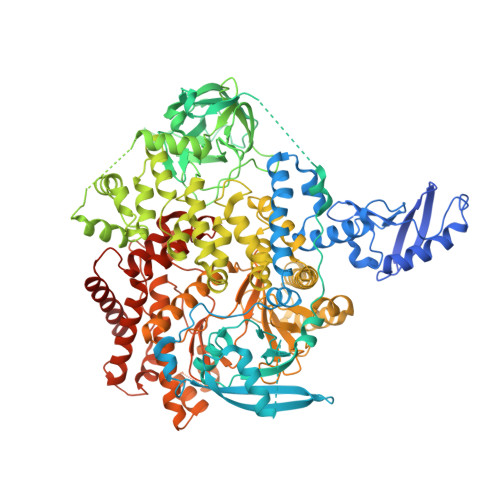
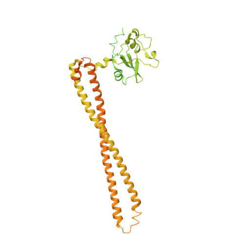
8GUB

Cryo-EM structure of cancer-specific PI3Kalpha mutant H1047R in complex with BYL-719


Experimental Data Snapshot

  • Method: ELECTRON MICROSCOPY
  • Resolution: 2.73 Å
  • Aggregation State: PARTICLE 
  • Reconstruction Method: SINGLE PARTICLE 

Starting Model: experimental
View more details

wwPDB Validation   3D Report Full Report


This is version 1.1 of the entry. See complete history


Literature

Cryo-EM structures of cancer-specific helical and kinase domain mutations of PI3K alpha.

Liu, X.Zhou, Q.Hart, J.R.Xu, Y.Yang, S.Yang, D.Vogt, P.K.Wang, M.W.

(2022) Proc Natl Acad Sci U S A 119: e2215621119-e2215621119

  • DOI: https://doi.org/10.1073/pnas.2215621119
  • Primary Citation of Related Structures:  
    8GUA, 8GUB, 8GUD

  • PubMed Abstract: 

    Phosphoinositide 3-kinases (PI3Ks) are a family of lipid kinases that perform multiple and important cellular functions. The protein investigated here belongs to class IA of the PI3Ks; it is a dimer consisting of a catalytic subunit, p110α, and a regulatory subunit, p85α, and is referred to as PI3Kα. The catalytic subunit p110α is frequently mutated in cancer. The mutations induce a gain of function and constitute a driving force in cancer development. About 80% of these mutations lead to single-amino-acid substitutions in one of three sites of p110α: two in the helical domain of the protein (E542K and E545K) and one at the C-terminus of the kinase domain (H1047R). Here, we report the cryo-electron microscopy structures of these mutants in complex with the p110α-specific inhibitor BYL-719. The H1047R mutant rotates its sidechain to a new position and weakens the kα11 activation loop interaction, thereby reducing the inhibitory effect of p85α on p110α. E542K and E545K completely abolish the tight interaction between the helical domain of p110α and the N-terminal SH2 domain of p85α and lead to the disruption of all p85α binding and a dramatic increase in flexibility of the adaptor-binding domain (ABD) in p110α. Yet, the dimerization of PI3Kα is preserved through the ABD-p85α interaction. The local and global structural features induced by these mutations provide molecular insights into the activation of PI3Kα, deepen our understanding of the oncogenic mechanism of this important signaling molecule, and may facilitate the development of mutant-specific inhibitors.


  • Organizational Affiliation

    Department of Pharmacology, School of Basic Medical Sciences, Fudan University, Shanghai 200032, China.


Macromolecules
Find similar proteins by:  (by identity cutoff)  |  3D Structure
Entity ID: 1
MoleculeChains Sequence LengthOrganismDetailsImage
Phosphatidylinositol 4,5-bisphosphate 3-kinase catalytic subunit alpha isoform1,096Homo sapiensMutation(s): 1 
Gene Names: PIK3CA
EC: 2.7.1.137 (PDB Primary Data), 2.7.1.153 (PDB Primary Data), 2.7.11.1 (PDB Primary Data)
UniProt & NIH Common Fund Data Resources
Find proteins for P42336 (Homo sapiens)
Explore P42336 
Go to UniProtKB:  P42336
PHAROS:  P42336
GTEx:  ENSG00000121879 
Entity Groups  
Sequence Clusters30% Identity50% Identity70% Identity90% Identity95% Identity100% Identity
UniProt GroupP42336
Sequence Annotations
Expand
  • Reference Sequence
Find similar proteins by:  (by identity cutoff)  |  3D Structure
Entity ID: 2
MoleculeChains Sequence LengthOrganismDetailsImage
Phosphatidylinositol 3-kinase regulatory subunit alpha723Homo sapiensMutation(s): 0 
Gene Names: PIK3R1GRB1
UniProt & NIH Common Fund Data Resources
Find proteins for P27986 (Homo sapiens)
Explore P27986 
Go to UniProtKB:  P27986
PHAROS:  P27986
GTEx:  ENSG00000145675 
Entity Groups  
Sequence Clusters30% Identity50% Identity70% Identity90% Identity95% Identity100% Identity
UniProt GroupP27986
Sequence Annotations
Expand
  • Reference Sequence
Small Molecules
Ligands 1 Unique
IDChains Name / Formula / InChI Key2D Diagram3D Interactions
1LT (Subject of Investigation/LOI)
Query on 1LT

Download Ideal Coordinates CCD File 
C [auth A](2S)-N~1~-{4-methyl-5-[2-(1,1,1-trifluoro-2-methylpropan-2-yl)pyridin-4-yl]-1,3-thiazol-2-yl}pyrrolidine-1,2-dicarboxamide
C19 H22 F3 N5 O2 S
STUWGJZDJHPWGZ-LBPRGKRZSA-N
Experimental Data & Validation

Experimental Data

  • Method: ELECTRON MICROSCOPY
  • Resolution: 2.73 Å
  • Aggregation State: PARTICLE 
  • Reconstruction Method: SINGLE PARTICLE 
EM Software:
TaskSoftware PackageVersion
MODEL REFINEMENTPHENIX1.18.2-3874

Structure Validation

View Full Validation Report


Entry History & Funding Information

Deposition Data


Funding OrganizationLocationGrant Number
National Natural Science Foundation of China (NSFC)China81872915
National Natural Science Foundation of China (NSFC)China82073904
National Natural Science Foundation of China (NSFC)China82121005
National Natural Science Foundation of China (NSFC)China81973373
National Natural Science Foundation of China (NSFC)China21704064
Other government2018ZX09735-001
Other government2018ZX09711002-002-005
Other government2021ZD0203400
Other government2018YFA0507000
Other governmentZDKJ2021028
Other privateNNCAS-2017-1-CC
National Institutes of Health/National Cancer Institute (NIH/NCI)United StatesR35 CA197582
National Institutes of Health/National Cancer Institute (NIH/NCI)United StatesR50 CA243899

Revision History  (Full details and data files)

  • Version 1.0: 2022-11-23
    Type: Initial release
  • Version 1.1: 2024-07-03
    Changes: Data collection, Refinement description