8GSY

X-ray structure of Clostridium perfringens pili protein B N-terminal domain


Experimental Data Snapshot

  • Method: X-RAY DIFFRACTION
  • Resolution: 1.55 Å
  • R-Value Free: 0.240 
  • R-Value Work: 0.184 
  • R-Value Observed: 0.187 

wwPDB Validation   3D Report Full Report


This is version 1.1 of the entry. See complete history


Literature

Structural and biochemical characterization of Clostridium perfringens pili protein B collagen-binding domains.

Tamai, E.Yamada, M.Ishida, T.Arimura, N.Matsunami, R.Sekiya, H.Kamitori, S.

(2023) FEBS Lett 597: 1345-1354

  • DOI: https://doi.org/10.1002/1873-3468.14626
  • Primary Citation of Related Structures:  
    8GSX, 8GSY

  • PubMed Abstract: 

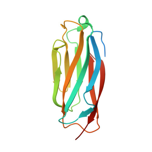
    Sortase-mediated pili are flexible rod proteins composed of major and minor/tip pilins, playing important roles in the initial adhesion of bacterial cells to host tissues. The pilus shaft is formed by covalent polymerization of major pilins, and the minor/tip pilin is covalently attached to the tip of the shaft involved in adhesion to the host cell. The Gram-positive bacterium Clostridium perfringens has a major pilin, and a minor/tip pilin (CppB) with the collagen-binding motif. Here, we report X-ray structures of CppB collagen-binding domains, collagen-binding assays and mutagenesis analysis, demonstrating that CppB collagen-binding domains adopt an L-shaped structure in open form, and that a small β-sheet unique to CppB provides a scaffold for a favourable binding site for collagen peptide.


  • Organizational Affiliation

    Department of Infectious Disease, College of Pharmaceutical Sciences, Matsuyama University, Japan.


Macromolecules
Find similar proteins by:  (by identity cutoff)  |  3D Structure
Entity ID: 1
MoleculeChains Sequence LengthOrganismDetailsImage
Peptidoglycan bound protein
A, B
144Clostridium perfringensMutation(s): 0 
Gene Names: NCTC8081_03081
UniProt
Find proteins for A0A2X3IRB0 (Clostridium perfringens)
Explore A0A2X3IRB0 
Go to UniProtKB:  A0A2X3IRB0
Entity Groups  
Sequence Clusters30% Identity50% Identity70% Identity90% Identity95% Identity100% Identity
UniProt GroupA0A2X3IRB0
Sequence Annotations
Expand
  • Reference Sequence
Experimental Data & Validation

Experimental Data

  • Method: X-RAY DIFFRACTION
  • Resolution: 1.55 Å
  • R-Value Free: 0.240 
  • R-Value Work: 0.184 
  • R-Value Observed: 0.187 
  • Space Group: P 32 2 1
Unit Cell:
Length ( Å )Angle ( ˚ )
a = 39.08α = 90
b = 39.08β = 90
c = 290.59γ = 120
Software Package:
Software NamePurpose
REFMACrefinement
XDSdata reduction
XSCALEdata scaling
MOLREPphasing

Structure Validation

View Full Validation Report


Entry History & Funding Information

Deposition Data


Funding OrganizationLocationGrant Number
Japan Society for the Promotion of Science (JSPS)Japan18K06087

Revision History  (Full details and data files)

  • Version 1.0: 2023-06-07
    Type: Initial release
  • Version 1.1: 2023-11-29
    Changes: Data collection, Refinement description