8GN9 | pdb_00008gn9

SPFH domain of Pyrococcus horikoshii stomatin


Experimental Data Snapshot

  • Method: X-RAY DIFFRACTION
  • Resolution: 2.50 Å
  • R-Value Free: 
    0.278 (Depositor), 0.274 (DCC) 
  • R-Value Work: 
    0.215 (Depositor), 0.214 (DCC) 
  • R-Value Observed: 
    0.222 (Depositor) 

Starting Model: experimental
View more details

wwPDB Validation 3D Report Full Report

Validation slider image for 8GN9

This is version 1.1 of the entry. See complete history

Literature

Structural and mutational studies suggest key residues to determine whether stomatin SPFH domains form dimers or trimers.

Komatsu, T.Matsui, I.Yokoyama, H.

(2022) Biochem Biophys Rep 32: 101384-101384

  • DOI: https://doi.org/10.1016/j.bbrep.2022.101384
  • Primary Citation Related Structures: 
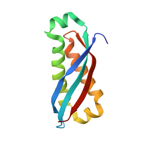
    8GN9

  • PubMed Abstract: 

    Stomatin is a major integral membrane protein in human erythrocytes. In a form of hemolytic anemia known as hereditary stomatocytosis, stomatin is deficient in the erythrocyte membrane due to mis-trafficking. It is a member of stomatin, prohibitin, flotillin, and HflK/C (SPFH) domain proteins, and SPFH proteins could function as membrane-bound oligomeric scaffolding proteins in lipid rafts. The previously determined structure of the SPFH domain of Pyrococcus horikoshii (Ph) stomatin formed a trimer, whereas that of mouse stomatin formed a dimer. To elucidate the difference of oligomerization state, structural and chromatographic analyses using Ph stomatin were performed, and the key residues were suggested to determine whether SPFH domains form dimers or trimers. From gel-filtration analyses, PhStom (56-234) formed a trimer or tetramer, whereas PhStom (63-234) and PhStom (56-234) K59S formed a dimer. The residues 56-62, particularly Lys59, were involved in trimerization. Based on the crystal structure of PhStom (63-234), it formed a banana-shaped dimer, as observed in mouse stomatin. Thus, residues 162-168 are involved in dimerization. This study provides important insight into the molecular function and oligomerization state of stomatin.


  • Organizational Affiliation
    • Faculty of Pharmaceutical Sciences, Tokyo University of Science, 2641 Yamazaki, Noda, Chiba, 278-8510, Japan.

Macromolecule Content 

  • Total Structure Weight: 12.31 kDa 
  • Atom Count: 862 
  • Modeled Residue Count: 106 
  • Deposited Residue Count: 109 
  • Unique protein chains: 1

Macromolecules

Find similar proteins by:|  3D Structure
Entity ID: 1
MoleculeChains  Sequence LengthOrganismDetailsImage
Stomatin homolog PH1511109Pyrococcus horikoshiiMutation(s): 0 
Gene Names: PH1511
UniProt
Find proteins for O59180 (Pyrococcus horikoshii (strain ATCC 700860 / DSM 12428 / JCM 9974 / NBRC 100139 / OT-3))
Explore O59180 
Go to UniProtKB:  O59180
Entity Groups
Sequence Clusters30% Identity50% Identity70% Identity90% Identity95% Identity100% Identity
UniProt GroupO59180
Sequence Annotations
Expand
Reference Sequence

Small Molecules

Ligands 1 Unique
IDChains Name / Formula / InChI Key2D Diagram3D Interactions
NA

Query on NA


Download:Ideal Coordinates CCD File
B [auth A]SODIUM ION
Na
FKNQFGJONOIPTF-UHFFFAOYSA-N

Experimental Data & Validation

Experimental Data

  • Method: X-RAY DIFFRACTION
  • Resolution: 2.50 Å
  • R-Value Free:  0.278 (Depositor), 0.274 (DCC) 
  • R-Value Work:  0.215 (Depositor), 0.214 (DCC) 
  • R-Value Observed: 0.222 (Depositor) 
Space Group: P 41 21 2
Unit Cell:
Length ( Å )Angle ( ˚ )
a = 28.014α = 90
b = 28.014β = 90
c = 240.63γ = 90
Software Package:
Software NamePurpose
REFMACrefinement
PDB_EXTRACTdata extraction
XDSdata reduction
SCALAdata scaling
MOLREPphasing

Structure Validation

View Full Validation Report


Entry History 

& Funding Information

Deposition Data


Funding OrganizationLocationGrant Number
Japan Society for the Promotion of Science (JSPS)Japan17K07316

Revision History  (Full details and data files)

  • Version 1.0: 2022-12-07
    Type: Initial release
  • Version 1.1: 2023-11-29
    Changes: Data collection, Refinement description