8GL3 | pdb_00008gl3

De novo design of monomeric helical bundles for pH-controlled membrane lysis


Experimental Data Snapshot

  • Method: X-RAY DIFFRACTION
  • Resolution: 2.30 Å
  • R-Value Free: 
    0.284 (Depositor), 0.283 (DCC) 
  • R-Value Work: 
    0.238 (Depositor), 0.238 (DCC) 
  • R-Value Observed: 
    0.240 (Depositor) 

Starting Model: in silico
View more details

wwPDB Validation 3D Report Full Report

Validation slider image for 8GL3

This is version 1.2 of the entry. See complete history

Literature

De novo design of monomeric helical bundles for pH-controlled membrane lysis.

Goldbach, N.Benna, I.Wicky, B.I.M.Croft, J.T.Carter, L.Bera, A.K.Nguyen, H.Kang, A.Sankaran, B.Yang, E.C.Lee, K.K.Baker, D.

(2023) Protein Sci 32: e4769-e4769

  • DOI: https://doi.org/10.1002/pro.4769
  • Primary Citation Related Structures: 
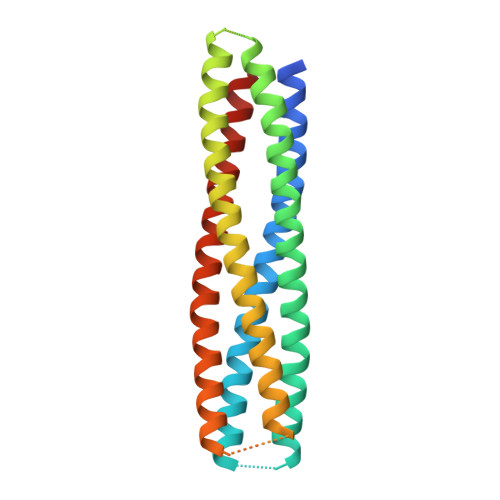
    8GL3

  • PubMed Abstract: 

    Targeted intracellular delivery via receptor-mediated endocytosis requires the delivered cargo to escape the endosome to prevent lysosomal degradation. This can in principle be achieved by membrane lysis tightly restricted to endosomal membranes upon internalization to avoid general membrane insertion and lysis. Here, we describe the design of small monomeric proteins with buried histidine containing pH-responsive hydrogen bond networks and membrane permeating amphipathic helices. Of the 30 designs that were experimentally tested, all expressed in Escherichia coli, 13 were monomeric with the expected secondary structure, and 4 designs disrupted artificial liposomes in a pH-dependent manner. Mutational analysis showed that the buried histidine hydrogen bond networks mediate pH-responsiveness and control lysis of model membranes within a very narrow range of pH (6.0-5.5) with almost no lysis occurring at neutral pH. These tightly controlled lytic monomers could help mediate endosomal escape in designed targeted delivery platforms.


  • Organizational Affiliation
    • Institute for Protein Design, University of Washington, Seattle, Washington, USA.

Macromolecule Content 

  • Total Structure Weight: 27.6 kDa 
  • Atom Count: 1,631 
  • Modeled Residue Count: 189 
  • Deposited Residue Count: 232 
  • Unique protein chains: 1

Macromolecules

Find similar proteins by:|  3D Structure
Entity ID: 1
MoleculeChains  Sequence LengthOrganismDetailsImage
pRLB-519232synthetic constructMutation(s): 0 

Experimental Data & Validation

Experimental Data

  • Method: X-RAY DIFFRACTION
  • Resolution: 2.30 Å
  • R-Value Free:  0.284 (Depositor), 0.283 (DCC) 
  • R-Value Work:  0.238 (Depositor), 0.238 (DCC) 
  • R-Value Observed: 0.240 (Depositor) 
Space Group: I 1 2 1
Unit Cell:
Length ( Å )Angle ( ˚ )
a = 32.12α = 90
b = 44.703β = 90.43
c = 182.579γ = 90
Software Package:
Software NamePurpose
PHENIXrefinement
DIALSdata reduction
DIALSdata scaling
PHASERphasing

Structure Validation

View Full Validation Report


Entry History 

& Funding Information

Deposition Data


Funding OrganizationLocationGrant Number
National Science Foundation (NSF, United States)United States--
Howard Hughes Medical Institute (HHMI)United States--

Revision History  (Full details and data files)

  • Version 1.0: 2023-09-06
    Type: Initial release
  • Version 1.1: 2023-09-20
    Changes: Refinement description
  • Version 1.2: 2023-11-08
    Changes: Database references