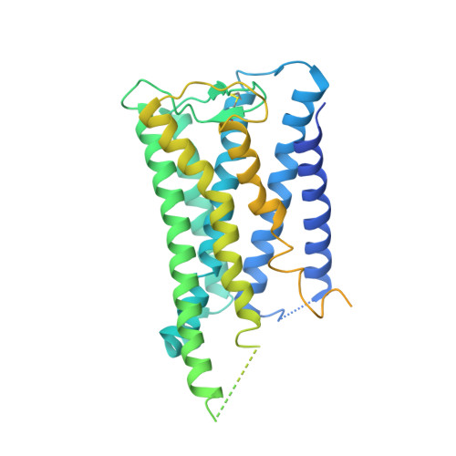
8GCP

Cryo-EM Structure of the Prostaglandin E2 Receptor 4 Coupled to G Protein


Experimental Data Snapshot

  • Method: ELECTRON MICROSCOPY
  • Resolution: 3.10 Å
  • Aggregation State: PARTICLE 
  • Reconstruction Method: SINGLE PARTICLE 

wwPDB Validation   3D Report Full Report


This is version 1.2 of the entry. See complete history


Literature

Single hormone or synthetic agonist induces G s /G i coupling selectivity of EP receptors via distinct binding modes and propagating paths.

Huang, S.M.Xiong, M.Y.Liu, L.Mu, J.Wang, M.W.Jia, Y.L.Cai, K.Tie, L.Zhang, C.Cao, S.Wen, X.Wang, J.L.Guo, S.C.Li, Y.Qu, C.X.He, Q.T.Cai, B.Y.Xue, C.Gan, S.Xie, Y.Cong, X.Yang, Z.Kong, W.Li, S.Li, Z.Xiao, P.Yang, F.Yu, X.Guan, Y.F.Zhang, X.Liu, Z.Yang, B.X.Du, Y.Sun, J.P.

(2023) Proc Natl Acad Sci U S A 120: e2216329120-e2216329120

  • DOI: https://doi.org/10.1073/pnas.2216329120
  • Primary Citation of Related Structures:  
    8GCM, 8GCP, 8GD9, 8GDA, 8GDB, 8GDC

  • PubMed Abstract: 

    To accomplish concerted physiological reactions, nature has diversified functions of a single hormone at at least two primary levels: 1) Different receptors recognize the same hormone, and 2) different cellular effectors couple to the same hormone-receptor pair [R.P. Xiao, Sci STKE 2001 , re15 (2001); L. Hein, J. D. Altman, B.K. Kobilka, Nature 402 , 181-184 (1999); Y. Daaka, L. M. Luttrell, R. J. Lefkowitz, Nature 390 , 88-91 (1997)]. Not only these questions lie in the heart of hormone actions and receptor signaling but also dissecting mechanisms underlying these questions could offer therapeutic routes for refractory diseases, such as kidney injury (KI) or X-linked nephrogenic diabetes insipidus (NDI). Here, we identified that G s -biased signaling, but not G i activation downstream of EP4, showed beneficial effects for both KI and NDI treatments. Notably, by solving Cryo-electron microscope (cryo-EM) structures of EP3-G i , EP4-G s , and EP4-G i in complex with endogenous prostaglandin E 2 (PGE 2 )or two synthetic agonists and comparing with PGE 2 -EP2-G s structures, we found that unique primary sequences of prostaglandin E2 receptor (EP) receptors and distinct conformational states of the EP4 ligand pocket govern the G s /G i transducer coupling selectivity through different structural propagation paths, especially via TM6 and TM7, to generate selective cytoplasmic structural features. In particular, the orientation of the PGE 2 ω-chain and two distinct pockets encompassing agonist L902688 of EP4 were differentiated by their G s /G i coupling ability. Further, we identified common and distinct features of cytoplasmic side of EP receptors for G s /G i coupling and provide a structural basis for selective and biased agonist design of EP4 with therapeutic potential.


  • Organizational Affiliation

    Department of Physiology and Pathophysiology, School of Basic Medical Sciences, State Key Laboratory of Vascular Homeostasis and Remodeling, Peking University, Beijing 100191, China.


Macromolecules
Find similar proteins by:  (by identity cutoff)  |  3D Structure
Entity ID: 1
MoleculeChains Sequence LengthOrganismDetailsImage
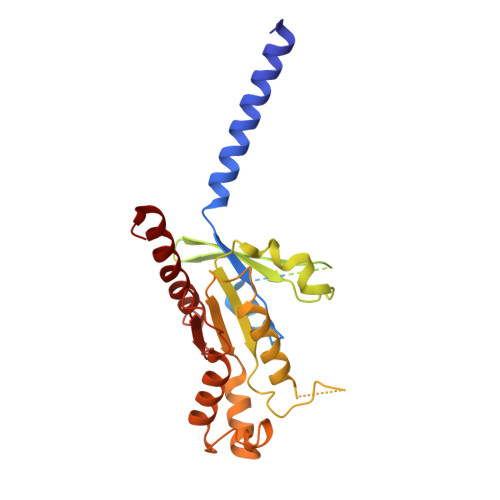
Guanine nucleotide-binding protein G(i) subunit alpha-1354Homo sapiensMutation(s): 0 
Gene Names: GNAI1
EC: 3.6.5
Membrane Entity: Yes 
UniProt & NIH Common Fund Data Resources
Find proteins for P63096 (Homo sapiens)
Explore P63096 
Go to UniProtKB:  P63096
PHAROS:  P63096
GTEx:  ENSG00000127955 
Entity Groups  
Sequence Clusters30% Identity50% Identity70% Identity90% Identity95% Identity100% Identity
UniProt GroupP63096
Sequence Annotations
Expand
  • Reference Sequence
Find similar proteins by:  (by identity cutoff)  |  3D Structure
Entity ID: 2
MoleculeChains Sequence LengthOrganismDetailsImage
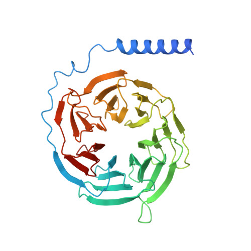
Guanine nucleotide-binding protein G(I)/G(S)/G(T) subunit beta-1358Homo sapiensMutation(s): 0 
Gene Names: GNB1
UniProt & NIH Common Fund Data Resources
Find proteins for P62873 (Homo sapiens)
Explore P62873 
Go to UniProtKB:  P62873
PHAROS:  P62873
GTEx:  ENSG00000078369 
Entity Groups  
Sequence Clusters30% Identity50% Identity70% Identity90% Identity95% Identity100% Identity
UniProt GroupP62873
Sequence Annotations
Expand
  • Reference Sequence
Find similar proteins by:  (by identity cutoff)  |  3D Structure
Entity ID: 3
MoleculeChains Sequence LengthOrganismDetailsImage
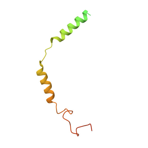
Guanine nucleotide-binding protein subunit gamma108Homo sapiensMutation(s): 0 
Gene Names: mRhiFer1_005810
Membrane Entity: Yes 
UniProt & NIH Common Fund Data Resources
Find proteins for P59768 (Homo sapiens)
Explore P59768 
Go to UniProtKB:  P59768
PHAROS:  P59768
GTEx:  ENSG00000186469 
Entity Groups  
Sequence Clusters30% Identity50% Identity70% Identity90% Identity95% Identity100% Identity
UniProt GroupP59768
Sequence Annotations
Expand
  • Reference Sequence
Find similar proteins by:  (by identity cutoff)  |  3D Structure
Entity ID: 4
MoleculeChains Sequence LengthOrganismDetailsImage
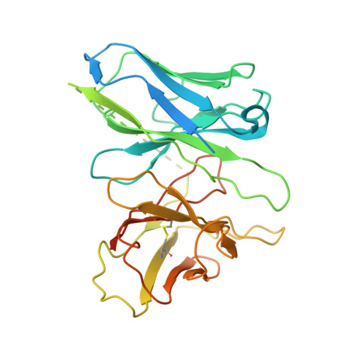
scFvD [auth E]307Homo sapiensMutation(s): 0 
Entity Groups  
Sequence Clusters30% Identity50% Identity70% Identity90% Identity95% Identity100% Identity
Sequence Annotations
Expand
  • Reference Sequence
Find similar proteins by:  (by identity cutoff)  |  3D Structure
Entity ID: 5
MoleculeChains Sequence LengthOrganismDetailsImage
Prostaglandin E2 receptor EP4 subtypeE [auth R]488Homo sapiensMutation(s): 0 
Gene Names: PTGER4PTGER2
Membrane Entity: Yes 
UniProt & NIH Common Fund Data Resources
Find proteins for P35408 (Homo sapiens)
Explore P35408 
Go to UniProtKB:  P35408
PHAROS:  P35408
GTEx:  ENSG00000171522 
Entity Groups  
Sequence Clusters30% Identity50% Identity70% Identity90% Identity95% Identity100% Identity
UniProt GroupP35408
Sequence Annotations
Expand
  • Reference Sequence
Small Molecules
Ligands 1 Unique
IDChains Name / Formula / InChI Key2D Diagram3D Interactions
P2E (Subject of Investigation/LOI)
Query on P2E

Download Ideal Coordinates CCD File 
F [auth R](Z)-7-[(1R,2R,3R)-3-hydroxy-2-[(E,3S)-3-hydroxyoct-1-enyl]-5-oxo-cyclopentyl]hept-5-enoic acid
C20 H32 O5
XEYBRNLFEZDVAW-ARSRFYASSA-N
Binding Affinity Annotations 
IDSourceBinding Affinity
P2E BindingDB:  8GCP Ki: min: 0.11, max: 3.1 (nM) from 5 assay(s)
IC50: min: 0.55, max: 9.9 (nM) from 5 assay(s)
EC50: min: 0.02, max: 7.5 (nM) from 6 assay(s)
Experimental Data & Validation

Experimental Data

  • Method: ELECTRON MICROSCOPY
  • Resolution: 3.10 Å
  • Aggregation State: PARTICLE 
  • Reconstruction Method: SINGLE PARTICLE 

Structure Validation

View Full Validation Report


Entry History & Funding Information

Deposition Data


Funding OrganizationLocationGrant Number
National Natural Science Foundation of China (NSFC)China81825022

Revision History  (Full details and data files)

  • Version 1.0: 2024-01-03
    Type: Initial release
  • Version 1.1: 2024-01-10
    Changes: Database references
  • Version 1.2: 2024-11-06
    Changes: Data collection, Structure summary