8G6P | pdb_00008g6p

Crystal structure of Mycobacterium thermoresistibile MurE in complex with ADP and 2,6-Diaminopimelic acid


Experimental Data Snapshot

  • Method: X-RAY DIFFRACTION
  • Resolution: 1.45 Å
  • R-Value Free: 
    0.190 (Depositor), 0.173 (DCC) 
  • R-Value Work: 
    0.170 (Depositor), 0.171 (DCC) 
  • R-Value Observed: 
    0.171 (Depositor) 

Starting Model: experimental
View more details

wwPDB Validation 3D Report Full Report

Validation slider image for 8G6P

Ligand Structure Quality Assessment 


This is version 2.0 of the entry. See complete history

Literature

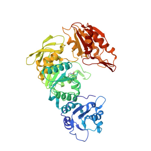
The crystal structure of Mycobacterium thermoresistibile MurE ligase reveals the binding mode of the substrate m-diaminopimelate.

Rossini, N.O.Silva, C.Dias, M.V.B.

(2023) J Struct Biol 215: 107957-107957

  • DOI: https://doi.org/10.1016/j.jsb.2023.107957
  • Primary Citation Related Structures: 
    8G6P

  • PubMed Abstract: 

    The cytoplasmatic biosynthesis of the stem peptide from the peptidoglycan in bacteria involves six steps, which have the role of three ATP-dependent Mur ligases that incorporate three consecutive amino acids to a substrate precursor. MurE is the last Mur ligase to incorporate a free amino acid. Although the structure of MurE from Mycobacterium tuberculosis (MtbMurE) was determined at 3.0 Å, the binding mode of meso-Diaminopimelate (m-DAP) and the effect of substrate absence is unknown. Herein, we show the structure of MurE from M. thermoresistibile (MthMurE) in complex with ADP and m-DAP at 1.4 Å resolution. The analysis of the structure indicates key conformational changes that the substrate UDP-MurNAc-L-Ala-D-Glu (UAG) and the free amino acid m-DAP cause on the MthMurE conformation. We observed several movements of domains or loop regions that displace their position in order to perform enzymatic catalysis. Since MthMurE has a high similarity to MtbMurE, this enzyme could also guide strategies for structure-based antimicrobial discovery to fight against tuberculosis or other mycobacterial infections.


  • Organizational Affiliation
    • Department of Microbiology, Institute of Biomedical Science, University of São Paulo, Av. Prof Lineu Prestes, 1374, CEP 05508-000 São Paulo, SP, Brazil.

Macromolecule Content 

  • Total Structure Weight: 54.11 kDa 
  • Atom Count: 4,510 
  • Modeled Residue Count: 508 
  • Deposited Residue Count: 508 
  • Unique protein chains: 1

Macromolecules

Find similar proteins by:|  3D Structure
Entity ID: 1
MoleculeChains  Sequence LengthOrganismDetailsImage
UDP-N-acetylmuramoyl-L-alanyl-D-glutamate--2,6-diaminopimelate ligase508Mycolicibacterium thermoresistibileMutation(s): 0 
Gene Names: murE
EC: 6.3.2.13
UniProt
Find proteins for G7CMG5 (Mycolicibacterium thermoresistibile (strain ATCC 19527 / DSM 44167 / CIP 105390 / JCM 6362 / NCTC 10409 / 316))
Explore G7CMG5 
Go to UniProtKB:  G7CMG5
Entity Groups
Sequence Clusters30% Identity50% Identity70% Identity90% Identity95% Identity100% Identity
UniProt GroupG7CMG5
Sequence Annotations
Expand
Reference Sequence

Small Molecules

Ligands 4 Unique
IDChains Name / Formula / InChI Key2D Diagram3D Interactions
ADP
(Subject of Investigation/LOI)

Query on ADP


Download:Ideal Coordinates CCD File
E [auth A]ADENOSINE-5'-DIPHOSPHATE
C10 H15 N5 O10 P2
XTWYTFMLZFPYCI-KQYNXXCUSA-N
API
(Subject of Investigation/LOI)

Query on API


Download:Ideal Coordinates CCD File
J [auth A]2,6-DIAMINOPIMELIC ACID
C7 H14 N2 O4
GMKMEZVLHJARHF-SYDPRGILSA-N
GOL

Query on GOL


Download:Ideal Coordinates CCD File
C [auth A]
D [auth A]
F [auth A]
G [auth A]
H [auth A]
C [auth A],
D [auth A],
F [auth A],
G [auth A],
H [auth A],
I [auth A]
GLYCEROL
C3 H8 O3
PEDCQBHIVMGVHV-UHFFFAOYSA-N
MG
(Subject of Investigation/LOI)

Query on MG


Download:Ideal Coordinates CCD File
B [auth A],
K [auth A],
L [auth A]
MAGNESIUM ION
Mg
JLVVSXFLKOJNIY-UHFFFAOYSA-N
Modified Residues  1 Unique
IDChains TypeFormula2D DiagramParent
KCX
Query on KCX
A
L-PEPTIDE LINKINGC7 H14 N2 O4LYS

Experimental Data & Validation

Experimental Data

  • Method: X-RAY DIFFRACTION
  • Resolution: 1.45 Å
  • R-Value Free:  0.190 (Depositor), 0.173 (DCC) 
  • R-Value Work:  0.170 (Depositor), 0.171 (DCC) 
  • R-Value Observed: 0.171 (Depositor) 
Space Group: P 43 21 2
Unit Cell:
Length ( Å )Angle ( ˚ )
a = 62.792α = 90
b = 62.792β = 90
c = 238.267γ = 90
Software Package:
Software NamePurpose
PHENIXrefinement
Aimlessdata scaling
XDSdata reduction
MOLREPphasing
PDB_EXTRACTdata extraction

Structure Validation

View Full Validation Report


Ligand Structure Quality Assessment 


Entry History 

& Funding Information

Deposition Data


Funding OrganizationLocationGrant Number
Sao Paulo Research Foundation (FAPESP)Brazil--

Revision History  (Full details and data files)

  • Version 1.0: 2023-04-05
    Type: Initial release
  • Version 1.1: 2023-07-19
    Changes: Data collection
  • Version 2.0: 2023-11-15
    Changes: Atomic model, Data collection