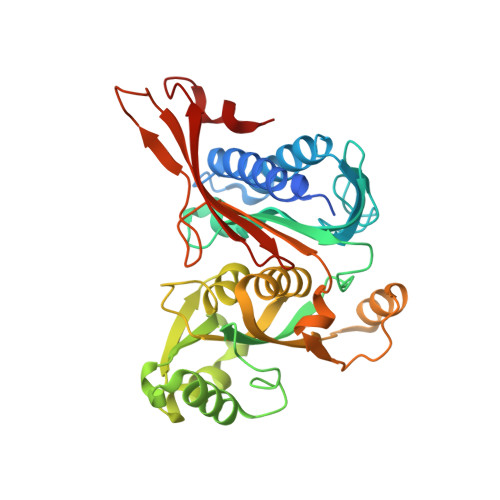
8G5W

Structure of the Class II Fructose-1,6-Bisphophatase from Francisella tularensis complexed with native metal cofactor Mn++


Experimental Data Snapshot

  • Method: X-RAY DIFFRACTION
  • Resolution: 2.00 Å
  • R-Value Free: 0.204 
  • R-Value Work: 0.161 
  • R-Value Observed: 0.163 

Starting Model: experimental
View more details

wwPDB Validation   3D Report Full Report


Ligand Structure Quality Assessment 


This is version 1.2 of the entry. See complete history


Literature

New structures of Class II Fructose-1,6-Bisphosphatase from Francisella tularensis provide a framework for a novel catalytic mechanism for the entire class.

Selezneva, A.I.Harding, L.N.M.Gutka, H.J.Movahedzadeh, F.Abad-Zapatero, C.

(2023) PLoS One 18: e0274723-e0274723

  • DOI: https://doi.org/10.1371/journal.pone.0274723
  • Primary Citation of Related Structures:  
    7TXA, 7TXB, 7TXG, 8G5W, 8G5X

  • PubMed Abstract: 

    Class II Fructose-1,6-bisphosphatases (FBPaseII) (EC: 3.1.3.11) are highly conserved essential enzymes in the gluconeogenic pathway of microorganisms. Previous crystallographic studies of FBPasesII provided insights into various inactivated states of the enzyme in different species. Presented here is the first crystal structure of FBPaseII in an active state, solved for the enzyme from Francisella tularensis (FtFBPaseII), containing native metal cofactor Mn2+ and complexed with catalytic product fructose-6-phosphate (F6P). Another crystal structure of the same enzyme complex is presented in the inactivated state due to the structural changes introduced by crystal packing. Analysis of the interatomic distances among the substrate, product, and divalent metal cations in the catalytic centers of the enzyme led to a revision of the catalytic mechanism suggested previously for class II FBPases. We propose that phosphate-1 is cleaved from the substrate fructose-1,6-bisphosphate (F1,6BP) by T89 in a proximal α-helix backbone (G88-T89-T90-I91-T92-S93-K94) in which the substrate transition state is stabilized by the positive dipole of the 〈-helix backbone. Once cleaved a water molecule found in the active site liberates the inorganic phosphate from T89 completing the catalytic mechanism. Additionally, a crystal structure of Mycobacterium tuberculosis FBPaseII (MtFBPaseII) containing a bound F1,6BP is presented to further support the substrate binding and novel catalytic mechanism suggested for this class of enzymes.


  • Organizational Affiliation

    Institute for Tuberculosis Research, University of Illinois at Chicago, Chicago, Illinois, United States of America.


Macromolecules
Find similar proteins by:  (by identity cutoff)  |  3D Structure
Entity ID: 1
MoleculeChains Sequence LengthOrganismDetailsImage
Fructose-1,6-bisphosphatase
A, B, C, D
348Francisella cf. tularensis subsp. novicida 3523Mutation(s): 0 
Gene Names: glpXFNC33_05530FNC42_07040
UniProt
Find proteins for Q5NEJ8 (Francisella tularensis subsp. tularensis (strain SCHU S4 / Schu 4))
Explore Q5NEJ8 
Go to UniProtKB:  Q5NEJ8
Entity Groups  
Sequence Clusters30% Identity50% Identity70% Identity90% Identity95% Identity100% Identity
UniProt GroupQ5NEJ8
Sequence Annotations
Expand
  • Reference Sequence
Small Molecules
Ligands 2 Unique
IDChains Name / Formula / InChI Key2D Diagram3D Interactions
GOL (Subject of Investigation/LOI)
Query on GOL

Download Ideal Coordinates CCD File 
E [auth A]
F [auth A]
G [auth A]
H [auth A]
I [auth A]
E [auth A],
F [auth A],
G [auth A],
H [auth A],
I [auth A],
K [auth B],
L [auth B],
M [auth B],
N [auth B],
P [auth C],
Q [auth C],
R [auth C],
S [auth C],
U [auth D],
V [auth D],
W [auth D],
X [auth D],
Y [auth D]
GLYCEROL
C3 H8 O3
PEDCQBHIVMGVHV-UHFFFAOYSA-N
MN (Subject of Investigation/LOI)
Query on MN

Download Ideal Coordinates CCD File 
J [auth A],
O [auth B],
T [auth C],
Z [auth D]
MANGANESE (II) ION
Mn
WAEMQWOKJMHJLA-UHFFFAOYSA-N
Experimental Data & Validation

Experimental Data

  • Method: X-RAY DIFFRACTION
  • Resolution: 2.00 Å
  • R-Value Free: 0.204 
  • R-Value Work: 0.161 
  • R-Value Observed: 0.163 
  • Space Group: P 1
Unit Cell:
Length ( Å )Angle ( ˚ )
a = 63.845α = 67.831
b = 75.879β = 68.136
c = 77.616γ = 76.372
Software Package:
Software NamePurpose
PHENIXrefinement
HKL-2000data reduction
SCALEPACKdata scaling
MOLREPphasing
REFMACrefinement

Structure Validation

View Full Validation Report


Ligand Structure Quality Assessment 


Entry History & Funding Information

Deposition Data


Funding OrganizationLocationGrant Number
Other privatePotts Memorial Foundation G3541
Other privateChicago Biomedical Consortium 084679-00001

Revision History  (Full details and data files)

  • Version 1.0: 2023-06-28
    Type: Initial release
  • Version 1.1: 2023-07-05
    Changes: Database references
  • Version 1.2: 2024-05-22
    Changes: Data collection