8G4U | pdb_00008g4u

Final ketosynthase+acyltransferase of the erythromycin modular polyketide synthase


Experimental Data Snapshot

  • Method: X-RAY DIFFRACTION
  • Resolution: 2.90 Å
  • R-Value Free: 
    0.314 (Depositor), 0.315 (DCC) 
  • R-Value Work: 
    0.239 (Depositor), 0.239 (DCC) 
  • R-Value Observed: 
    0.240 (Depositor) 

Starting Model: experimental
View more details

wwPDB Validation 3D Report Full Report

Validation slider image for 8G4U

This is version 1.1 of the entry. See complete history

Literature

Crystal structures reveal the framework of cis -acyltransferase modular polyketide synthases.

Keatinge-Clay, A.T.Miyazawa, T.Zhang, J.Ray, K.A.Lutgens, J.D.Bista, R.Lin, S.N.

(2023) bioRxiv 

  • DOI: https://doi.org/10.1101/2023.02.11.528132
  • Primary Citation Related Structures: 
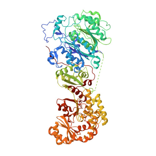
    8G4U

  • PubMed Abstract: 

    Although the domains of cis -acyltransferase ( cis -AT) modular polyketide synthases (PKS's) have been understood at atomic resolution for over a decade, the domain-domain interactions responsible for the architectures and activities of these giant molecular assembly lines remain largely uncharacterized. The multimeric structure of the α 6 β 6 fungal fatty acid synthase (FAS) provides 6 equivalent reaction chambers for its acyl carrier protein (ACP) domains to shuttle carbon building blocks and the growing acyl chain between surrounding, oriented enzymatic domains. The presumed homodimeric oligomerization of cis -AT assembly lines is insufficient to provide similar reaction chambers; however, the crystal structure of a ketosynthase (KS)+AT didomain presented here and three already reported show an interaction between the AT domains appropriate for lateral multimerization. This interaction was used to construct a framework for the pikromycin PKS from its KS, AT, and docking domains that contains highly-ordered reaction chambers. Its AT domains also mediate vertical interactions, both with upstream KS domains and downstream docking domains.

Macromolecule Content 

  • Total Structure Weight: 363.82 kDa 
  • Atom Count: 22,987 
  • Modeled Residue Count: 3,169 
  • Deposited Residue Count: 3,524 
  • Unique protein chains: 1

Macromolecules

Find similar proteins by:|  3D Structure
Entity ID: 1
MoleculeChains  Sequence LengthOrganismDetailsImage
EryAIII
A, B, C, D
881Saccharopolyspora erythraeaMutation(s): 1 
Gene Names: eryAIII
EC: 2.3.1.94
UniProt
Find proteins for Q03133 (Saccharopolyspora erythraea)
Explore Q03133 
Go to UniProtKB:  Q03133
Entity Groups
Sequence Clusters30% Identity50% Identity70% Identity90% Identity95% Identity100% Identity
UniProt GroupQ03133
Sequence Annotations
Expand
Reference Sequence

Experimental Data & Validation

Experimental Data

  • Method: X-RAY DIFFRACTION
  • Resolution: 2.90 Å
  • R-Value Free:  0.314 (Depositor), 0.315 (DCC) 
  • R-Value Work:  0.239 (Depositor), 0.239 (DCC) 
  • R-Value Observed: 0.240 (Depositor) 
Space Group: P 1 21 1
Unit Cell:
Length ( Å )Angle ( ˚ )
a = 71.21α = 90
b = 167.94β = 99.43
c = 184.23γ = 90
Software Package:
Software NamePurpose
PHENIXrefinement
XDSdata reduction
XDSdata scaling
PHENIXphasing

Structure Validation

View Full Validation Report


Entry History 

& Funding Information

Deposition Data


Funding OrganizationLocationGrant Number
National Institutes of Health/National Institute of General Medical Sciences (NIH/NIGMS)United States106112

Revision History  (Full details and data files)

  • Version 1.0: 2023-03-01
    Type: Initial release
  • Version 1.1: 2024-05-22
    Changes: Data collection