8FYN | pdb_00008fyn

MicroED structure of A2A from plasma milled lamellae


Experimental Data Snapshot

  • Method: ELECTRON CRYSTALLOGRAPHY
  • Resolution: 2.00 Å
  • R-Value Free: 
    0.279 (Depositor) 
  • R-Value Work: 
    0.220 (Depositor) 
  • R-Value Observed: 
    0.223 (Depositor) 

wwPDB Validation 3D Report Full Report

Validation slider image for 8FYN

This is version 1.2 of the entry. See complete history

Literature

A robust approach for MicroED sample preparation of lipidic cubic phase embedded membrane protein crystals.

Martynowycz, M.W.Shiriaeva, A.Clabbers, M.T.B.Nicolas, W.J.Weaver, S.J.Hattne, J.Gonen, T.

(2023) Nat Commun 14: 1086-1086

  • DOI: https://doi.org/10.1038/s41467-023-36733-4
  • Primary Citation Related Structures: 
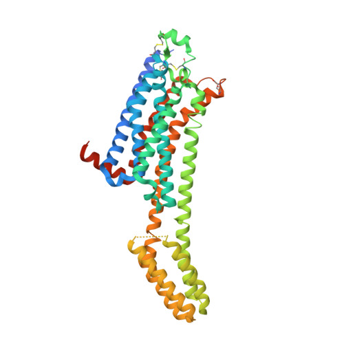
    8FYN, 8FYO, 8FYP, 8FYQ, 8FYR, 8FYS

  • PubMed Abstract: 

    Crystallizing G protein-coupled receptors (GPCRs) in lipidic cubic phase (LCP) often yields crystals suited for the cryogenic electron microscopy (cryoEM) method microcrystal electron diffraction (MicroED). However, sample preparation is challenging. Embedded crystals cannot be targeted topologically. Here, we use an integrated fluorescence light microscope (iFLM) inside of a focused ion beam and scanning electron microscope (FIB-SEM) to identify fluorescently labeled GPCR crystals. Crystals are targeted using the iFLM and LCP is milled using a plasma focused ion beam (pFIB). The optimal ion source for preparing biological lamellae is identified using standard crystals of proteinase K. Lamellae prepared using either argon or xenon produced the highest quality data and structures. MicroED data are collected from the milled lamellae and the structures are determined. This study outlines a robust approach to identify and mill membrane protein crystals for MicroED and demonstrates plasma ion-beam milling is a powerful tool for preparing biological lamellae.


  • Organizational Affiliation
    • Howard Hughes Medical Institute, University of California, Los Angeles, CA, 90095, USA.

Macromolecule Content 

  • Total Structure Weight: 57.87 kDa 
  • Atom Count: 3,622 
  • Modeled Residue Count: 390 
  • Deposited Residue Count: 447 
  • Unique protein chains: 1

Macromolecules

Find similar proteins by:|  3D Structure
Entity ID: 1
MoleculeChains  Sequence LengthOrganismDetailsImage
Adenosine receptor A2a,Soluble cytochrome b562447Homo sapiensMutation(s): 0 
Gene Names: ADORA2AADORA2cybC
Membrane Entity: Yes 
UniProt & NIH Common Fund Data Resources
Find proteins for P0ABE7 (Escherichia coli)
Explore P0ABE7 
Go to UniProtKB:  P0ABE7
Find proteins for P29274 (Homo sapiens)
Explore P29274 
Go to UniProtKB:  P29274
PHAROS:  P29274
GTEx:  ENSG00000128271 
Entity Groups
Sequence Clusters30% Identity50% Identity70% Identity90% Identity95% Identity100% Identity
UniProt GroupsP0ABE7P29274
Sequence Annotations
Expand
Reference Sequence

Small Molecules

Ligands 6 Unique
IDChains Name / Formula / InChI Key2D Diagram3D Interactions
CLR

Query on CLR


Download:Ideal Coordinates CCD File
C [auth A],
D [auth A],
E [auth A]
CHOLESTEROL
C27 H46 O
HVYWMOMLDIMFJA-DPAQBDIFSA-N
OLB

Query on OLB


Download:Ideal Coordinates CCD File
X [auth A](2S)-2,3-dihydroxypropyl (9Z)-octadec-9-enoate
C21 H40 O4
RZRNAYUHWVFMIP-QJRAZLAKSA-N
OLC

Query on OLC


Download:Ideal Coordinates CCD File
U [auth A],
V [auth A],
W [auth A],
Y [auth A],
Z [auth A]
(2R)-2,3-dihydroxypropyl (9Z)-octadec-9-enoate
C21 H40 O4
RZRNAYUHWVFMIP-GDCKJWNLSA-N
ZMA

Query on ZMA


Download:Ideal Coordinates CCD File
B [auth A]4-{2-[(7-amino-2-furan-2-yl[1,2,4]triazolo[1,5-a][1,3,5]triazin-5-yl)amino]ethyl}phenol
C16 H15 N7 O2
PWTBZOIUWZOPFT-UHFFFAOYSA-N
OLA

Query on OLA


Download:Ideal Coordinates CCD File
F [auth A]
G [auth A]
H [auth A]
I [auth A]
J [auth A]
F [auth A],
G [auth A],
H [auth A],
I [auth A],
J [auth A],
K [auth A],
L [auth A],
M [auth A],
N [auth A],
O [auth A],
P [auth A],
Q [auth A],
R [auth A],
S [auth A],
T [auth A]
OLEIC ACID
C18 H34 O2
ZQPPMHVWECSIRJ-KTKRTIGZSA-N
NA

Query on NA


Download:Ideal Coordinates CCD File
AA [auth A]SODIUM ION
Na
FKNQFGJONOIPTF-UHFFFAOYSA-N

Experimental Data & Validation

Experimental Data

  • Method: ELECTRON CRYSTALLOGRAPHY
  • Resolution: 2.00 Å
  • R-Value Free:  0.279 (Depositor) 
  • R-Value Work:  0.220 (Depositor) 
  • R-Value Observed: 0.223 (Depositor) 
Space Group: C 2 2 21
Unit Cell:
Length ( Å )Angle ( ˚ )
a = 39.08α = 90
b = 177.51β = 90
c = 139.29γ = 90
Software Package:
Software NamePurpose
phenix.refinerefinement
PHENIXrefinement

Structure Validation

View Full Validation Report


Entry History 

& Funding Information

Deposition Data


Funding OrganizationLocationGrant Number
Howard Hughes Medical Institute (HHMI)United States--
National Institutes of Health/National Institute of General Medical Sciences (NIH/NIGMS)United StatesP41GM136508

Revision History  (Full details and data files)

  • Version 1.0: 2023-03-08
    Type: Initial release
  • Version 1.1: 2023-03-22
    Changes: Source and taxonomy
  • Version 1.2: 2024-10-23
    Changes: Data collection, Structure summary