8FR5 | pdb_00008fr5

Crystal structure of the Human Smacovirus 1 Rep domain


Experimental Data Snapshot

  • Method: X-RAY DIFFRACTION
  • Resolution: 1.33 Å
  • R-Value Free: 
    0.224 (Depositor), 0.225 (DCC) 
  • R-Value Work: 
    0.187 (Depositor), 0.187 (DCC) 
  • R-Value Observed: 
    0.191 (Depositor) 

Starting Model: in silico
View more details

wwPDB Validation 3D Report Full Report

Validation slider image for 8FR5

This is version 1.0 of the entry. See complete history

Literature

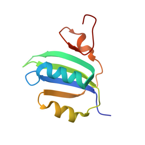
The crystal structure of the human smacovirus 1 Rep domain.

Limon, L.K.Shi, K.Dao, A.Rugloski, J.Tompkins, K.J.Aihara, H.Gordon, W.R.Evans 3rd, R.L.

(2023) Acta Crystallogr F Struct Biol Commun 79: 295-300

  • DOI: https://doi.org/10.1107/S2053230X23009536
  • Primary Citation Related Structures: 
    8FR5

  • PubMed Abstract: 

    Replication initiator proteins (Reps) from the HUH endonuclease family process specific single-stranded DNA sequences to initiate rolling-circle replication in viruses. Here, the first crystal structure of the apo state of a Rep domain from the smacovirus family is reported. The structure of the human smacovirus 1 Rep domain was obtained at 1.33 Å resolution and represents an expansion of the HUH endonuclease superfamily, allowing greater diversity in bioconjugation-tag applications.


  • Organizational Affiliation
    • Department of Biochemistry, Molecular Biology and Biophysics, University of Minnesota, Minneapolis, MN 55455, USA.

Macromolecule Content 

  • Total Structure Weight: 11.63 kDa 
  • Atom Count: 784 
  • Modeled Residue Count: 92 
  • Deposited Residue Count: 97 
  • Unique protein chains: 1

Macromolecules

Find similar proteins by:|  3D Structure
Entity ID: 1
MoleculeChains  Sequence LengthOrganismDetailsImage
Rep97Human smacovirus 1Mutation(s): 1 
Gene Names: Rep
UniProt
Find proteins for A0ACD6B8S6 (Human smacovirus 1)
Explore A0ACD6B8S6 
Go to UniProtKB:  A0ACD6B8S6
Entity Groups
Sequence Clusters30% Identity50% Identity70% Identity90% Identity95% Identity100% Identity
UniProt GroupA0ACD6B8S6
Sequence Annotations
Expand
Reference Sequence

Experimental Data & Validation

Experimental Data

  • Method: X-RAY DIFFRACTION
  • Resolution: 1.33 Å
  • R-Value Free:  0.224 (Depositor), 0.225 (DCC) 
  • R-Value Work:  0.187 (Depositor), 0.187 (DCC) 
  • R-Value Observed: 0.191 (Depositor) 
Space Group: P 1 21 1
Unit Cell:
Length ( Å )Angle ( ˚ )
a = 31.16α = 90
b = 49.37β = 110.301
c = 31.38γ = 90
Software Package:
Software NamePurpose
PHENIXrefinement
HKL-2000data reduction
HKL-2000data scaling
PHASERphasing

Structure Validation

View Full Validation Report


Entry History 

& Funding Information

Deposition Data


Funding OrganizationLocationGrant Number
National Institutes of Health/National Institute of General Medical Sciences (NIH/NIGMS)United StatesR35 GM119483

Revision History  (Full details and data files)

  • Version 1.0: 2023-12-27
    Type: Initial release