8FMI | pdb_00008fmi

Crystal structure of human KRAS at 1.12 A


Experimental Data Snapshot

  • Method: X-RAY DIFFRACTION
  • Resolution: 1.12 Å
  • R-Value Free: 
    0.147 (Depositor), 0.147 (DCC) 
  • R-Value Work: 
    0.119 (Depositor), 0.120 (DCC) 
  • R-Value Observed: 
    0.121 (Depositor) 

Starting Model: experimental
View more details

wwPDB Validation 3D Report Full Report

Validation slider image for 8FMI

Ligand Structure Quality Assessment 


This is version 1.1 of the entry. See complete history

Literature

Crystal Packing Reveals a Potential Autoinhibited KRAS Dimer Interface and a Strategy for Small-Molecule Inhibition of RAS Signaling.

Brenner, R.J.Landgraf, A.D.Bum-Erdene, K.Gonzalez-Gutierrez, G.Meroueh, S.O.

(2023) Biochemistry 62: 3206-3213

  • DOI: https://doi.org/10.1021/acs.biochem.3c00378
  • Primary Citation Related Structures: 
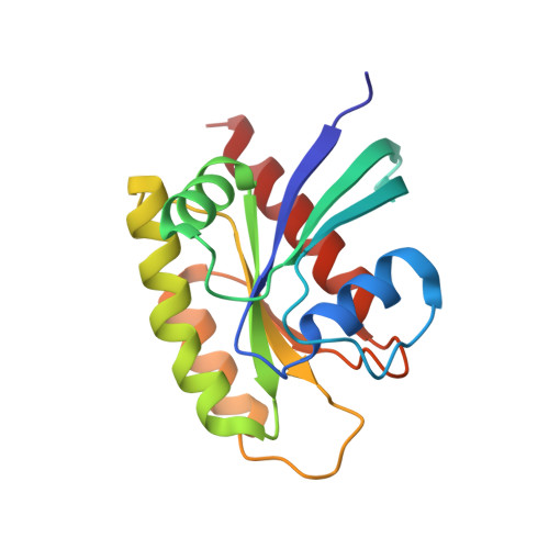
    8FMI, 8FMJ, 8FMK

  • PubMed Abstract: 

    KRAS GTPases harbor oncogenic mutations in more than 25% of human tumors. KRAS is considered to be largely undruggable due to the lack of a suitable small-molecule binding site. Here, we report a unique crystal structure of His-tagged KRAS G12D that reveals a remarkable conformational change. The Switch I loop of one His-KRAS G12D structure extends into the Switch I/II pocket of another His-KRAS G12D in an adjacent unit cell to create an elaborate interface that is reminiscent of high-affinity protein-protein complexes. We explore the contributions of amino acids at this interface using alanine-scanning studies with alchemical free energy perturbation calculations based on explicit-solvent molecular dynamics simulations. Several interface amino acids were found to be hot spots as they contributed more than 1.5 kcal/mol to the protein-protein interaction. Computational analysis of the complex revealed the presence of two large binding pockets that possess physicochemical features typically found in pockets considered druggable. Small-molecule binding to these pockets may stabilize this autoinhibited structure of KRAS if it exists in cells to provide a new strategy to inhibit RAS signaling.


  • Organizational Affiliation
    • Department of Biochemistry and Molecular Biology, Indiana University School of Medicine, Indianapolis, Indiana 46202, United States.

Macromolecule Content 

  • Total Structure Weight: 19.78 kDa 
  • Atom Count: 1,717 
  • Modeled Residue Count: 170 
  • Deposited Residue Count: 170 
  • Unique protein chains: 1

Macromolecules

Find similar proteins by:|  3D Structure
Entity ID: 1
MoleculeChains  Sequence LengthOrganismDetailsImage
Isoform 2B of GTPase KRas170Homo sapiensMutation(s): 1 
Gene Names: KRASKRAS2RASK2
EC: 3.6.5.2
UniProt & NIH Common Fund Data Resources
Find proteins for P01116 (Homo sapiens)
Explore P01116 
Go to UniProtKB:  P01116
PHAROS:  P01116
GTEx:  ENSG00000133703 
Entity Groups
Sequence Clusters30% Identity50% Identity70% Identity90% Identity95% Identity100% Identity
UniProt GroupP01116
Sequence Annotations
Expand
Reference Sequence

Small Molecules

Experimental Data & Validation

Experimental Data

  • Method: X-RAY DIFFRACTION
  • Resolution: 1.12 Å
  • R-Value Free:  0.147 (Depositor), 0.147 (DCC) 
  • R-Value Work:  0.119 (Depositor), 0.120 (DCC) 
  • R-Value Observed: 0.121 (Depositor) 
Space Group: P 21 21 21
Unit Cell:
Length ( Å )Angle ( ˚ )
a = 38.93α = 90
b = 39.81β = 90
c = 91.67γ = 90
Software Package:
Software NamePurpose
PHENIXrefinement
XDSdata reduction
Aimlessdata scaling
PHASERphasing

Structure Validation

View Full Validation Report


Ligand Structure Quality Assessment 


Entry History 

& Funding Information

Deposition Data


Funding OrganizationLocationGrant Number
National Institutes of Health/National Cancer Institute (NIH/NCI)United StatesR01CA264471
National Institutes of Health/National Cancer Institute (NIH/NCI)United StatesR01CA197928

Revision History  (Full details and data files)

  • Version 1.0: 2023-11-29
    Type: Initial release
  • Version 1.1: 2023-12-06
    Changes: Database references