8FJ9 | pdb_00008fj9

Structure of the catalytic domain of Streptococcus mutans GtfB in tetragonal space group P4322


Experimental Data Snapshot

  • Method: X-RAY DIFFRACTION
  • Resolution: 2.50 Å
  • R-Value Free: 
    0.241 (Depositor), 0.243 (DCC) 
  • R-Value Work: 
    0.212 (Depositor), 0.216 (DCC) 
  • R-Value Observed: 
    0.214 (Depositor) 

Starting Model: experimental
View more details

wwPDB Validation 3D Report Full Report

Validation slider image for 8FJ9

Ligand Structure Quality Assessment 


This is version 1.1 of the entry. See complete history

Literature

The catalytic domains of Streptococcus mutans glucosyltransferases: a structural analysis.

Schormann, N.Patel, M.Thannickal, L.Purushotham, S.Wu, R.Mieher, J.L.Wu, H.Deivanayagam, C.

(2023) Acta Crystallogr F Struct Biol Commun 79: 119-127

  • DOI: https://doi.org/10.1107/S2053230X23003199
  • Primary Citation Related Structures: 
    8FJ9, 8FJC, 8FK4, 8FKL, 8FN5

  • PubMed Abstract: 

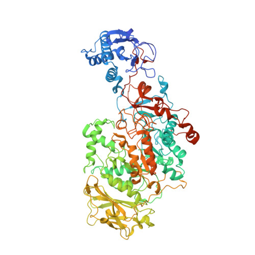
    Streptococcus mutans, found in the human oral cavity, is a significant contributor to the pathogenesis of dental caries. This bacterium expresses three genetically distinct types of glucosyltransferases named GtfB (GTF-I), GtfC (GTF-SI) and GtfD (GTF-S) that play critical roles in the development of dental plaque. The catalytic domains of GtfB, GtfC and GtfD contain conserved active-site residues for the overall enzymatic activity that relate to hydrolytic glycosidic cleavage of sucrose to glucose and fructose, release of fructose and generation of a glycosyl-enzyme intermediate in the reducing end. In a subsequent transglycosylation step, the glucosyl moiety is transferred to the nonreducing end of an acceptor to form a growing glucan polymer chain made up of glucose molecules. It has been proposed that both sucrose breakdown and glucan synthesis occur in the same active site of the catalytic domain, although the active site does not appear to be large enough to accommodate both functions. These three enzymes belong to glycoside hydrolase family 70 (GH70), which shows homology to glycoside hydrolase family 13 (GH13). GtfC synthesizes both soluble and insoluble glucans (α-1,3 and α-1,6 glycosidic linkages), while GtfB and GtfD synthesize only insoluble or soluble glucans, respectively. Here, crystal structures of the catalytic domains of GtfB and GtfD are reported. These structures are compared with previously determined structures of the catalytic domain of GtfC. With this work, apo structures and inhibitor-complex structures with acarbose are now available for the catalytic domains of GtfC and GtfB. The structure of GtfC with maltose allows further identification and comparison of active-site residues. A model of sucrose binding to GtfB is also included. The new structure of the catalytic domain of GtfD affords a structural comparison of the three S. mutans glycosyltransferases. Unfortunately, the catalytic domain of GtfD is not complete since crystallization resulted in the structure of a truncated protein lacking approximately 200 N-terminal residues of domain IV.


  • Organizational Affiliation
    • Department of Biochemistry and Molecular Genetics, University of Alabama at Birmingham, Birmingham, AL 35294, USA.

Macromolecule Content 

  • Total Structure Weight: 198.32 kDa 
  • Atom Count: 13,703 
  • Modeled Residue Count: 1,670 
  • Deposited Residue Count: 1,738 
  • Unique protein chains: 1

Macromolecules

Find similar proteins by:|  3D Structure
Entity ID: 1
MoleculeChains  Sequence LengthOrganismDetailsImage
Glucosyltransferase-I
A, B
869Streptococcus mutansMutation(s): 0 
Gene Names: gtfBSMU_1004
EC: 2.4.1.5
UniProt
Find proteins for P08987 (Streptococcus mutans serotype c (strain ATCC 700610 / UA159))
Explore P08987 
Go to UniProtKB:  P08987
Entity Groups
Sequence Clusters30% Identity50% Identity70% Identity90% Identity95% Identity100% Identity
UniProt GroupP08987
Sequence Annotations
Expand
Reference Sequence

Small Molecules

Ligands 4 Unique
IDChains Name / Formula / InChI Key2D Diagram3D Interactions
SO4
(Subject of Investigation/LOI)

Query on SO4


Download:Ideal Coordinates CCD File
BA [auth B]
CA [auth B]
DA [auth B]
EA [auth B]
FA [auth B]
BA [auth B],
CA [auth B],
DA [auth B],
EA [auth B],
FA [auth B],
G [auth A],
GA [auth B],
H [auth A],
HA [auth B],
I [auth A],
IA [auth B],
J [auth A],
JA [auth B],
K [auth A],
KA [auth B],
L [auth A],
LA [auth B],
M [auth A],
MA [auth B],
N [auth A],
NA [auth B],
O [auth A],
OA [auth B],
P [auth A],
Q [auth A],
R [auth A],
S [auth A],
T [auth A]
SULFATE ION
O4 S
QAOWNCQODCNURD-UHFFFAOYSA-L
EDO
(Subject of Investigation/LOI)

Query on EDO


Download:Ideal Coordinates CCD File
AA [auth B]
D [auth A]
E [auth A]
F [auth A]
W [auth B]
AA [auth B],
D [auth A],
E [auth A],
F [auth A],
W [auth B],
X [auth B],
Y [auth B],
Z [auth B]
1,2-ETHANEDIOL
C2 H6 O2
LYCAIKOWRPUZTN-UHFFFAOYSA-N
CA
(Subject of Investigation/LOI)

Query on CA


Download:Ideal Coordinates CCD File
C [auth A],
V [auth B]
CALCIUM ION
Ca
BHPQYMZQTOCNFJ-UHFFFAOYSA-N
CL
(Subject of Investigation/LOI)

Query on CL


Download:Ideal Coordinates CCD File
U [auth A]CHLORIDE ION
Cl
VEXZGXHMUGYJMC-UHFFFAOYSA-M

Experimental Data & Validation

Experimental Data

  • Method: X-RAY DIFFRACTION
  • Resolution: 2.50 Å
  • R-Value Free:  0.241 (Depositor), 0.243 (DCC) 
  • R-Value Work:  0.212 (Depositor), 0.216 (DCC) 
  • R-Value Observed: 0.214 (Depositor) 
Space Group: P 43 2 2
Unit Cell:
Length ( Å )Angle ( ˚ )
a = 150.13α = 90
b = 150.13β = 90
c = 302.783γ = 90
Software Package:
Software NamePurpose
REFMACrefinement
PDB_EXTRACTdata extraction
XDSdata reduction
Aimlessdata scaling
PHASERphasing

Structure Validation

View Full Validation Report


Ligand Structure Quality Assessment 


Entry History 

& Funding Information

Deposition Data


Funding OrganizationLocationGrant Number
Not funded--

Revision History  (Full details and data files)

  • Version 1.0: 2023-05-17
    Type: Initial release
  • Version 1.1: 2024-05-01
    Changes: Data collection, Database references