8FJ0

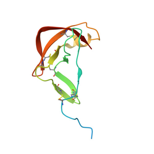
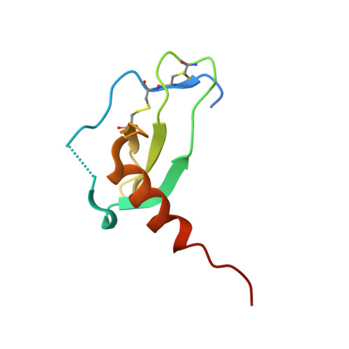
Crystal Structure of the Tick Evasin EVA-AAM1001(Y44A) Complexed to Human Chemokine CCL2


Experimental Data Snapshot

  • Method: X-RAY DIFFRACTION
  • Resolution: 2.91 Å
  • R-Value Free: 0.284 
  • R-Value Work: 0.254 
  • R-Value Observed: 0.255 

Starting Model: experimental
View more details

wwPDB Validation   3D Report Full Report


This is version 1.2 of the entry. See complete history


Literature

Engineering broad-spectrum inhibitors of inflammatory chemokines from subclass A3 tick evasins.

Devkota, S.R.Aryal, P.Pokhrel, R.Jiao, W.Perry, A.Panjikar, S.Payne, R.J.Wilce, M.C.J.Bhusal, R.P.Stone, M.J.

(2023) Nat Commun 14: 4204-4204

  • DOI: https://doi.org/10.1038/s41467-023-39879-3
  • Primary Citation of Related Structures:  
    7SCS, 7SCT, 7SCV, 8FJ0, 8FJ2, 8FJ3, 8FK6, 8FK8

  • PubMed Abstract: 

    Chemokines are key regulators of leukocyte trafficking and attractive targets for anti-inflammatory therapy. Evasins are chemokine-binding proteins from tick saliva, whose application as anti-inflammatory therapeutics will require manipulation of their chemokine target selectivity. Here we describe subclass A3 evasins, which are unique to the tick genus Amblyomma and distinguished from "classical" class A1 evasins by an additional disulfide bond near the chemokine recognition interface. The A3 evasin EVA-AAM1001 (EVA-A) bound to CC chemokines and inhibited their receptor activation. Unlike A1 evasins, EVA-A was not highly dependent on N- and C-terminal regions to differentiate chemokine targets. Structures of chemokine-bound EVA-A revealed a deep hydrophobic pocket, unique to A3 evasins, that interacts with the residue immediately following the CC motif of the chemokine. Mutations to this pocket altered the chemokine selectivity of EVA-A. Thus, class A3 evasins provide a suitable platform for engineering proteins with applications in research, diagnosis or anti-inflammatory therapy.


  • Organizational Affiliation

    Monash Biomedicine Discovery Institute, and Department of Biochemistry and Molecular Biology, Monash University, Clayton, VIC, 3800, Australia.


Macromolecules
Find similar proteins by:  (by identity cutoff)  |  3D Structure
Entity ID: 1
MoleculeChains Sequence LengthOrganismDetailsImage
Evasin P1243
A, B, C
103Amblyomma americanumMutation(s): 1 
UniProt
Find proteins for A0A0C9S461 (Amblyomma americanum)
Explore A0A0C9S461 
Go to UniProtKB:  A0A0C9S461
Entity Groups  
Sequence Clusters30% Identity50% Identity70% Identity90% Identity95% Identity100% Identity
UniProt GroupA0A0C9S461
Sequence Annotations
Expand
  • Reference Sequence
Find similar proteins by:  (by identity cutoff)  |  3D Structure
Entity ID: 2
MoleculeChains Sequence LengthOrganismDetailsImage
C-C motif chemokine 2D,
E [auth F],
F [auth E]
76Homo sapiensMutation(s): 0 
Gene Names: CCL2MCP1SCYA2
UniProt & NIH Common Fund Data Resources
Find proteins for P13500 (Homo sapiens)
Explore P13500 
Go to UniProtKB:  P13500
PHAROS:  P13500
GTEx:  ENSG00000108691 
Entity Groups  
Sequence Clusters30% Identity50% Identity70% Identity90% Identity95% Identity100% Identity
UniProt GroupP13500
Sequence Annotations
Expand
  • Reference Sequence
Experimental Data & Validation

Experimental Data

  • Method: X-RAY DIFFRACTION
  • Resolution: 2.91 Å
  • R-Value Free: 0.284 
  • R-Value Work: 0.254 
  • R-Value Observed: 0.255 
  • Space Group: P 1 21 1
Unit Cell:
Length ( Å )Angle ( ˚ )
a = 82.446α = 90
b = 49.636β = 112.91
c = 88.319γ = 90
Software Package:
Software NamePurpose
PHENIXrefinement
PHENIXrefinement
XDSdata reduction
Aimlessdata scaling
PHASERphasing

Structure Validation

View Full Validation Report


Entry History & Funding Information

Deposition Data


Funding OrganizationLocationGrant Number
National Health and Medical Research Council (NHMRC, Australia)AustraliaAPP1140867

Revision History  (Full details and data files)

  • Version 1.0: 2023-03-29
    Type: Initial release
  • Version 1.1: 2023-07-26
    Changes: Database references
  • Version 1.2: 2024-10-16
    Changes: Data collection, Structure summary