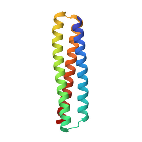
8FIH

Multi-state design of two-state switchable hinge proteins


Experimental Data Snapshot

  • Method: X-RAY DIFFRACTION
  • Resolution: 2.20 Å
  • R-Value Free: 0.248 
  • R-Value Work: 0.191 
  • R-Value Observed: 0.195 

Starting Model: in silico
View more details

wwPDB Validation   3D Report Full Report


Ligand Structure Quality Assessment 


This is version 1.2 of the entry. See complete history


Literature

Design of stimulus-responsive two-state hinge proteins.

Praetorius, F.Leung, P.J.Y.Tessmer, M.H.Broerman, A.Demakis, C.Dishman, A.F.Pillai, A.Idris, A.Juergens, D.Dauparas, J.Li, X.Levine, P.M.Lamb, M.Ballard, R.K.Gerben, S.R.Nguyen, H.Kang, A.Sankaran, B.Bera, A.K.Volkman, B.F.Nivala, J.Stoll, S.Baker, D.

(2023) Science 381: 754-760

  • DOI: https://doi.org/10.1126/science.adg7731
  • Primary Citation of Related Structures:  
    8FIH, 8FIN, 8FIQ, 8FIT, 8FVT

  • PubMed Abstract: 

    In nature, proteins that switch between two conformations in response to environmental stimuli structurally transduce biochemical information in a manner analogous to how transistors control information flow in computing devices. Designing proteins with two distinct but fully structured conformations is a challenge for protein design as it requires sculpting an energy landscape with two distinct minima. Here we describe the design of "hinge" proteins that populate one designed state in the absence of ligand and a second designed state in the presence of ligand. X-ray crystallography, electron microscopy, double electron-electron resonance spectroscopy, and binding measurements demonstrate that despite the significant structural differences the two states are designed with atomic level accuracy and that the conformational and binding equilibria are closely coupled.


  • Organizational Affiliation

    Department of Biochemistry, University of Washington, Seattle, WA, USA.


Macromolecules
Find similar proteins by:  (by identity cutoff)  |  3D Structure
Entity ID: 1
MoleculeChains Sequence LengthOrganismDetailsImage
3hb05
A, B, C
105synthetic constructMutation(s): 0 
Entity Groups  
Sequence Clusters30% Identity50% Identity70% Identity90% Identity95% Identity100% Identity
Sequence Annotations
Expand
  • Reference Sequence
Experimental Data & Validation

Experimental Data

  • Method: X-RAY DIFFRACTION
  • Resolution: 2.20 Å
  • R-Value Free: 0.248 
  • R-Value Work: 0.191 
  • R-Value Observed: 0.195 
  • Space Group: P 21 21 2
Unit Cell:
Length ( Å )Angle ( ˚ )
a = 66.018α = 90
b = 119.757β = 90
c = 38.8γ = 90
Software Package:
Software NamePurpose
PHENIXrefinement
XDSdata reduction
XSCALEdata scaling
PHASERphasing

Structure Validation

View Full Validation Report


Ligand Structure Quality Assessment 


Entry History & Funding Information

Deposition Data


Funding OrganizationLocationGrant Number
Howard Hughes Medical Institute (HHMI)United States--

Revision History  (Full details and data files)

  • Version 1.0: 2023-08-16
    Type: Initial release
  • Version 1.1: 2023-10-11
    Changes: Refinement description
  • Version 1.2: 2024-03-06
    Changes: Database references