8FHJ | pdb_00008fhj

Crystal structure of a FAD monooxygenease from Methylocystis sp. Strain SB2


Experimental Data Snapshot

  • Method: X-RAY DIFFRACTION
  • Resolution: 2.61 Å
  • R-Value Free: 
    0.254 (Depositor), 0.255 (DCC) 
  • R-Value Work: 
    0.204 (Depositor), 0.209 (DCC) 
  • R-Value Observed: 
    0.206 (Depositor) 

Starting Model: experimental
View more details

wwPDB Validation 3D Report Full Report

Validation slider image for 8FHJ

Ligand Structure Quality Assessment 


This is version 1.2 of the entry. See complete history

Literature

Crystal structure of MbnF: an NADPH-dependent flavin monooxygenase from Methylocystis strain SB2.

Stewart, A.Dershwitz, P.Stewart Jr., C.Sawaya, M.R.Yeates, T.O.Semrau, J.D.Zischka, H.DiSpirito, A.A.Bobik, T.A.

(2023) Acta Crystallogr F Struct Biol Commun 79: 111-118

  • DOI: https://doi.org/10.1107/S2053230X23003035
  • Primary Citation Related Structures: 
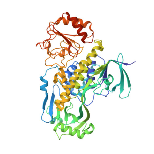
    8FHJ

  • PubMed Abstract: 

    Methanobactins (MBs) are ribosomally produced and post-translationally modified peptides (RiPPs) that are used by methanotrophs for copper acquisition. The signature post-translational modification of MBs is the formation of two heterocyclic groups, either an oxazolone, pyrazinedione or imidazolone group, with an associated thioamide from an X-Cys dipeptide. The precursor peptide (MbnA) for MB formation is found in a gene cluster of MB-associated genes. The exact biosynthetic pathway of MB formation is not yet fully understood, and there are still uncharacterized proteins in some MB gene clusters, particularly those that produce pyrazinedione or imidazolone rings. One such protein is MbnF, which is proposed to be a flavin monooxygenase (FMO) based on homology. To help to elucidate its possible function, MbnF from Methylocystis sp. strain SB2 was recombinantly produced in Escherichia coli and its X-ray crystal structure was resolved to 2.6 Å resolution. Based on its structural features, MbnF appears to be a type A FMO, most of which catalyze hydroxylation reactions. Preliminary functional characterization shows that MbnF preferentially oxidizes NADPH over NADH, supporting NAD(P)H-mediated flavin reduction, which is the initial step in the reaction cycle of several type A FMO enzymes. It is also shown that MbnF binds the precursor peptide for MB, with subsequent loss of the leader peptide sequence as well as the last three C-terminal amino acids, suggesting that MbnF might be needed for this process to occur. Finally, molecular-dynamics simulations revealed a channel in MbnF that is capable of accommodating the core MbnA fragment minus the three C-terminal amino acids.


  • Organizational Affiliation
    • Roy J. Carver Department of Biochemistry, Biophysics and Molecular Biology, Iowa State University, Ames, IA 50011-3260, USA.

Macromolecule Content 

  • Total Structure Weight: 185.13 kDa 
  • Atom Count: 12,534 
  • Modeled Residue Count: 1,594 
  • Deposited Residue Count: 1,671 
  • Unique protein chains: 1

Macromolecules

Find similar proteins by:|  3D Structure
Entity ID: 1
MoleculeChains  Sequence LengthOrganismDetailsImage
Monooxygenase
A, B, C
557Methylocystis sp. SB2Mutation(s): 0 
Gene Names: CTY30_04200
UniProt
Find proteins for A0ACD6BAX2 (Methylocystis sp. (strain SB2))
Explore A0ACD6BAX2 
Go to UniProtKB:  A0ACD6BAX2
Entity Groups
Sequence Clusters30% Identity50% Identity70% Identity90% Identity95% Identity100% Identity
UniProt GroupA0ACD6BAX2
Sequence Annotations
Expand
Reference Sequence

Small Molecules

Ligands 4 Unique
IDChains Name / Formula / InChI Key2D Diagram3D Interactions
FAD
(Subject of Investigation/LOI)

Query on FAD


Download:Ideal Coordinates CCD File
AA [auth C],
D [auth A],
L [auth B]
FLAVIN-ADENINE DINUCLEOTIDE
C27 H33 N9 O15 P2
VWWQXMAJTJZDQX-UYBVJOGSSA-N
GOL

Query on GOL


Download:Ideal Coordinates CCD File
BA [auth C]
E [auth A]
F [auth A]
M [auth B]
N [auth B]
BA [auth C],
E [auth A],
F [auth A],
M [auth B],
N [auth B],
O [auth B],
P [auth B],
Q [auth B],
R [auth B],
S [auth B]
GLYCEROL
C3 H8 O3
PEDCQBHIVMGVHV-UHFFFAOYSA-N
BR

Query on BR


Download:Ideal Coordinates CCD File
CA [auth C]
DA [auth C]
G [auth A]
H [auth A]
I [auth A]
CA [auth C],
DA [auth C],
G [auth A],
H [auth A],
I [auth A],
J [auth A],
K [auth A],
U [auth B],
V [auth B],
W [auth B],
X [auth B],
Y [auth B],
Z [auth B]
BROMIDE ION
Br
CPELXLSAUQHCOX-UHFFFAOYSA-M
NA

Query on NA


Download:Ideal Coordinates CCD File
T [auth B]SODIUM ION
Na
FKNQFGJONOIPTF-UHFFFAOYSA-N

Experimental Data & Validation

Experimental Data

  • Method: X-RAY DIFFRACTION
  • Resolution: 2.61 Å
  • R-Value Free:  0.254 (Depositor), 0.255 (DCC) 
  • R-Value Work:  0.204 (Depositor), 0.209 (DCC) 
  • R-Value Observed: 0.206 (Depositor) 
Space Group: C 1 2 1
Unit Cell:
Length ( Å )Angle ( ˚ )
a = 148.347α = 90
b = 96.765β = 116.41
c = 135.206γ = 90
Software Package:
Software NamePurpose
PHENIXrefinement
Aimlessdata scaling
PHASERphasing
autoPROCdata processing
XDSdata reduction

Structure Validation

View Full Validation Report


Ligand Structure Quality Assessment 


Entry History 

& Funding Information

Deposition Data


Funding OrganizationLocationGrant Number
Department of Energy (DOE, United States)United StatesDE-SC0018059
National Science Foundation (NSF, United States)United States1912482
Iowa State University (ISU) Bailey Research and Career Development AwardUnited StatesSG0600002

Revision History  (Full details and data files)

  • Version 1.0: 2023-05-03
    Type: Initial release
  • Version 1.1: 2023-05-31
    Changes: Database references, Refinement description
  • Version 1.2: 2024-05-22
    Changes: Data collection