8FFE | pdb_00008ffe

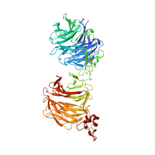
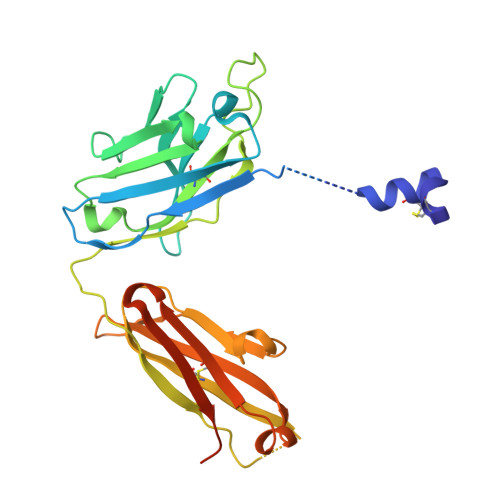
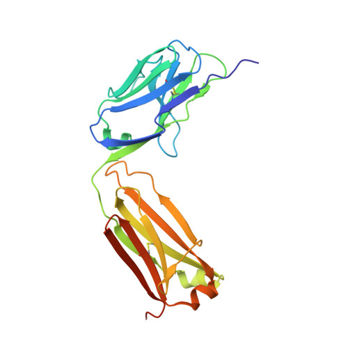
Crystal structure of LRP6 E1E2 domains bound to YW210.09 Fab and engineered XWnt8 peptide


Experimental Data Snapshot

  • Method: X-RAY DIFFRACTION
  • Resolution: 1.72 Å
  • R-Value Free: 
    0.231 (Depositor), 0.231 (DCC) 
  • R-Value Work: 
    0.185 (Depositor), 0.185 (DCC) 
  • R-Value Observed: 
    0.187 (Depositor) 

Starting Models: experimental
View more details

wwPDB Validation 3D Report Full Report

Validation slider image for 8FFE

This is version 1.2 of the entry. See complete history. 

Literature

Structure of the Wnt-Frizzled-LRP6 initiation complex reveals the basis for coreceptor discrimination.

Tsutsumi, N., Hwang, S., Waghray, D., Hansen, S., Jude, K.M., Wang, N., Miao, Y., Glassman, C.R., Caveney, N.A., Janda, C.Y., Hannoush, R.N., Garcia, K.C.

(2023) Proc Natl Acad Sci U S A 120: e2218238120-e2218238120

  • DOI: https://doi.org/10.1073/pnas.2218238120
  • Primary Citation Related Structures: 
    8CTG, 8FFE

  • PubMed Abstract: 

    Wnt morphogens are critical for embryonic development and tissue regeneration. Canonical Wnts form ternary receptor complexes composed of tissue-specific Frizzled (Fzd) receptors together with the shared LRP5/6 coreceptors to initiate β-catenin signaling. The cryo-EM structure of a ternary initiation complex of an affinity-matured XWnt8-Frizzled8-LRP6 complex elucidates the basis of coreceptor discrimination by canonical Wnts by means of their N termini and linker domains that engage the LRP6 E1E2 domain funnels. Chimeric Wnts bearing modular linker "grafts" were able to transfer LRP6 domain specificity between different Wnts and enable non-canonical Wnt5a to signal through the canonical pathway. Synthetic peptides comprising the linker domain serve as Wnt-specific antagonists. The structure of the ternary complex provides a topological blueprint for the orientation and proximity of Frizzled and LRP6 within the Wnt cell surface signalosome.


  • Organizational Affiliation: 
    • HHMI, Stanford University School of Medicine, Stanford, CA 94305.

Macromolecule Content 

  • Total Structure Weight: 125.4 kDa 
  • Atom Count: 9,116 
  • Modeled Residue Count: 1,067 
  • Deposited Residue Count: 1,114 
  • Unique protein chains: 3

Macromolecules

Find similar proteins by:|  3D Structure
Entity ID: 1
MoleculeChains  Sequence LengthOrganismDetailsImage
Low-density lipoprotein receptor-related protein 6613Homo sapiensMutation(s): 0 
Gene Names: LRP6
UniProt & NIH Common Fund Data Resources
Find proteins for O75581 (Homo sapiens)
Explore O75581 
Go to UniProtKB:  O75581
PHAROS:  O75581
GTEx:  ENSG00000070018 
Entity Groups
Sequence Clusters30% Identity50% Identity70% Identity90% Identity95% Identity100% Identity
UniProt GroupO75581
Glycosylation
Glycosylation Sites: 3Go to GlyGen: O75581-1
Sequence Annotations
Expand
Reference Sequence
Find similar proteins by:|  3D Structure
Entity ID: 2
MoleculeChains  Sequence LengthOrganismDetailsImage
YW210.09 Fab heavy chain with engineered XWnt8 NC peptide and linkerB [auth H]276Homo sapiensMutation(s): 0 
Entity Groups
Sequence Clusters30% Identity50% Identity70% Identity90% Identity95% Identity100% Identity
Sequence Annotations
Expand
Reference Sequence
Find similar proteins by:|  3D Structure
Entity ID: 3
MoleculeChains  Sequence LengthOrganismDetailsImage
YW210.09 Fab light chainC [auth L]225Homo sapiensMutation(s): 0 
Entity Groups
Sequence Clusters30% Identity50% Identity70% Identity90% Identity95% Identity100% Identity
Sequence Annotations
Expand
Reference Sequence

Oligosaccharides

Help  
Entity ID: 4
MoleculeChains Length2D Diagram GlycosylationD Interactions
alpha-L-fucopyranose-(1-3)-[2-acetamido-2-deoxy-beta-D-glucopyranose-(1-4)][alpha-L-fucopyranose-(1-6)]2-acetamido-2-deoxy-beta-D-glucopyranoseD [auth B]4N/AN-Glycosylation
Glycosylation Resources
GlyTouCan: G63564LA
GlyCosmos: G63564LA
GlyGen: G63564LA
Entity ID: 5
MoleculeChains Length2D Diagram GlycosylationD Interactions
beta-D-mannopyranose-(1-4)-2-acetamido-2-deoxy-beta-D-glucopyranose-(1-4)-[alpha-L-fucopyranose-(1-3)][alpha-L-fucopyranose-(1-6)]2-acetamido-2-deoxy-beta-D-glucopyranoseE [auth C]5N/AN-Glycosylation
Glycosylation Resources
GlyTouCan: G92953ND
GlyCosmos: G92953ND
GlyGen: G92953ND
Entity ID: 6
MoleculeChains Length2D Diagram GlycosylationD Interactions
alpha-D-mannopyranose-(1-3)-[2-acetamido-2-deoxy-beta-D-glucopyranose-(1-4)]2-acetamido-2-deoxy-beta-D-glucopyranoseF [auth D]3N/AN-Glycosylation

Small Molecules

Ligands 3 Unique
IDChains Name / Formula / InChI Key2D Diagram3D Interactions
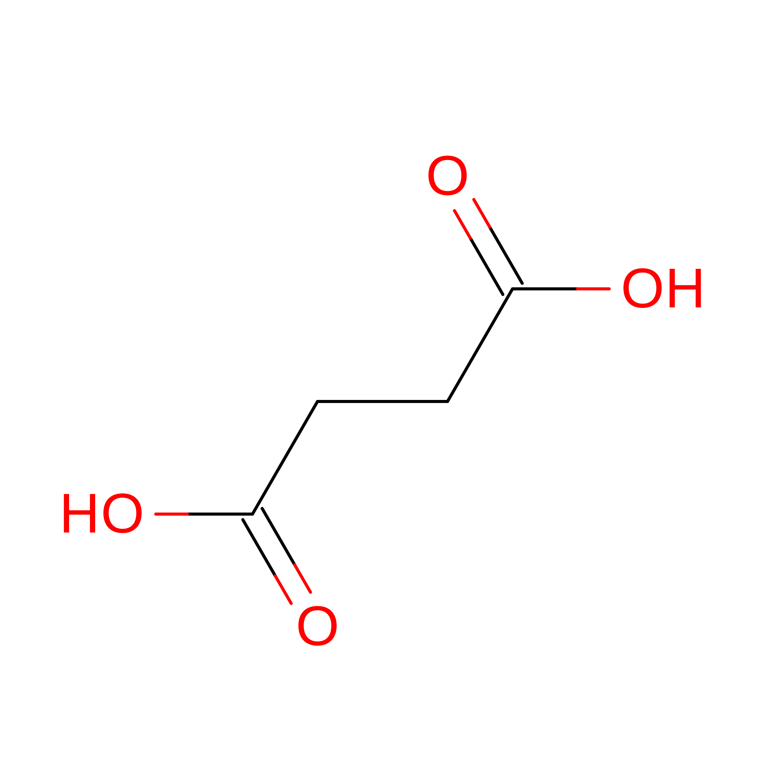
SIN

Query on SIN


Download:Ideal Coordinates CCD File
Q [auth L]SUCCINIC ACID
C4 H6 O4
KDYFGRWQOYBRFD-UHFFFAOYSA-N
GOL

Query on GOL


Download:Ideal Coordinates CCD File
G [auth A]
H [auth A]
I [auth A]
J [auth A]
N [auth A]
G [auth A],
H [auth A],
I [auth A],
J [auth A],
N [auth A],
O [auth H],
P [auth L]
GLYCEROL
C3 H8 O3
PEDCQBHIVMGVHV-UHFFFAOYSA-N
NA

Query on NA


Download:Ideal Coordinates CCD File
K [auth A],
L [auth A],
M [auth A]
SODIUM ION
Na
FKNQFGJONOIPTF-UHFFFAOYSA-N

Experimental Data & Validation

Experimental Data

  • Method: X-RAY DIFFRACTION
  • Resolution: 1.72 Å
  • R-Value Free:  0.231 (Depositor), 0.231 (DCC) 
  • R-Value Work:  0.185 (Depositor), 0.185 (DCC) 
  • R-Value Observed: 0.187 (Depositor) 
Space Group: C 1 2 1
Unit Cell:
Length ( Å )Angle ( ˚ )
a = 125.212α = 90
b = 91.385β = 103.67
c = 104.055γ = 90
Software Package:
Software NamePurpose
PHENIXrefinement
XDSdata reduction
XDSdata scaling
PHASERphasing

Structure Validation

View Full Validation Report


Entry History 

& Funding Information

Deposition Data


Funding OrganizationLocationGrant Number
National Institutes of Health/National Institute of Diabetes and Digestive and Kidney Disease (NIH/NIDDK)United StatesR01DK115728

Revision History  (Full details and data files)

  • Version 1.0: 2023-03-08
    Type: Initial release
  • Version 1.1: 2023-03-22
    Changes: Data collection, Database references
  • Version 1.2: 2024-11-20
    Changes: Data collection, Structure summary