8EY5

Human Serum Albumin with Cobalt (II) and Myristic Acid - crystal 3


Experimental Data Snapshot

  • Method: X-RAY DIFFRACTION
  • Resolution: 3.10 Å
  • R-Value Free: 0.261 
  • R-Value Work: 0.202 
  • R-Value Observed: 0.204 

wwPDB Validation   3D Report Full Report


Ligand Structure Quality Assessment 


This is version 2.3 of the entry. See complete history


Literature

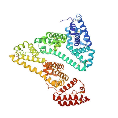
Structural and biochemical characterisation of Co2+-binding sites on serum albumins and their interplay with fatty acids

Wu, D.Gucwa, M.Czub, M.P.Cooper, D.R.Shabalin, I.G.Fritzen, R.Arya, S.Schwarz-Linek, U.Blindauer, C.A.Minor, W.Stewart, A.J.

(2023) Chem Sci 14: 6244-6258

  • DOI: https://doi.org/10.1039/d3sc01723k
  • Primary Citation of Related Structures:  
    8EW4, 8EW7, 8EY5

  • PubMed Abstract: 

    Serum albumin-Co 2+ interactions are of clinical importance. They play a role in mediating the physiological effects associated with cobalt toxicity and are central to the albumin cobalt binding (ACB) assay for diagnosis of myocardial ischemia. To further understand these processes, a deeper understanding of albumin-Co 2+ interactions is required. Here, we present the first crystallographic structures of human serum albumin (HSA; three structures) and equine serum albumin (ESA; one structure) in complex with Co 2+ . Amongst a total of sixteen sites bearing a cobalt ion across the structures, two locations were prominent, and they relate to metal-binding sites A and B. Site-directed mutagenesis and isothermal titration calorimetry (ITC) were employed to characterise sites on HSA. The results indicate that His9 and His67 contribute to the primary (putatively corresponding to site B) and secondary Co 2+ -binding sites (site A), respectively. The presence of additional multiple weak-affinity Co 2+ binding sites on HSA was also supported by ITC studies. Furthermore, addition of 5 molar equivalents of the non-esterified fatty acid palmitate (C16:0) reduced the Co 2+ -binding affinity at both sites A and B. The presence of bound myristate (C14:0) in the HSA crystal structures provided insight into the fatty acid-mediated structural changes that diminish the affinity of the protein toward Co 2+ . Together, these data provide further support for the idea that ischemia-modified albumin corresponds to albumin with excessive fatty-acid loading. Collectively, our findings provide a comprehensive understanding of the molecular underpinnings governing Co 2+ binding to serum albumin.


  • Organizational Affiliation

    School of Medicine, University of St Andrews St Andrews UK ajs21@st-andrews.ac.uk +44 (0)1334 463546.


Macromolecules
Find similar proteins by:  (by identity cutoff)  |  3D Structure
Entity ID: 1
MoleculeChains Sequence LengthOrganismDetailsImage
Serum albumin585Homo sapiensMutation(s): 0 
UniProt & NIH Common Fund Data Resources
Find proteins for P02768 (Homo sapiens)
Explore P02768 
Go to UniProtKB:  P02768
PHAROS:  P02768
GTEx:  ENSG00000163631 
Entity Groups  
Sequence Clusters30% Identity50% Identity70% Identity90% Identity95% Identity100% Identity
UniProt GroupP02768
Sequence Annotations
Expand
  • Reference Sequence
Small Molecules
Ligands 2 Unique
IDChains Name / Formula / InChI Key2D Diagram3D Interactions
MYR (Subject of Investigation/LOI)
Query on MYR

Download Ideal Coordinates CCD File 
G [auth A]
H [auth A]
I [auth A]
J [auth A]
K [auth A]
G [auth A],
H [auth A],
I [auth A],
J [auth A],
K [auth A],
L [auth A],
M [auth A],
N [auth A],
O [auth A]
MYRISTIC ACID
C14 H28 O2
TUNFSRHWOTWDNC-UHFFFAOYSA-N
CO (Subject of Investigation/LOI)
Query on CO

Download Ideal Coordinates CCD File 
B [auth A],
C [auth A],
D [auth A],
E [auth A],
F [auth A]
COBALT (II) ION
Co
XLJKHNWPARRRJB-UHFFFAOYSA-N
Experimental Data & Validation

Experimental Data

Unit Cell:
Length ( Å )Angle ( ˚ )
a = 185.966α = 90
b = 38.548β = 103.75
c = 93.409γ = 90
Software Package:
Software NamePurpose
REFMACrefinement
HKL-3000data reduction
HKL-3000data scaling
MOLREPphasing
PDB_EXTRACTdata extraction

Structure Validation

View Full Validation Report


Ligand Structure Quality Assessment 


Entry History & Funding Information

Deposition Data


Funding OrganizationLocationGrant Number
National Institutes of Health/National Institute of General Medical Sciences (NIH/NIGMS)United StatesGM132595

Revision History  (Full details and data files)

  • Version 1.0: 2022-11-09
    Type: Initial release
  • Version 1.1: 2023-02-01
    Changes: Other, Structure summary
  • Version 2.0: 2023-03-22
    Type: Coordinate replacement
    Reason: Polymer geometry
    Changes: Advisory, Atomic model, Data collection, Database references, Derived calculations, Refinement description, Structure summary
  • Version 2.1: 2023-04-19
    Changes: Author supporting evidence
  • Version 2.2: 2023-10-25
    Changes: Data collection, Refinement description
  • Version 2.3: 2024-06-26
    Changes: Database references