8EVN | pdb_00008evn

Sulfatase from Mycobacterium tuberculosis (Rv3406) in complex with N-oxalylglycine (NOG)


Experimental Data Snapshot

  • Method: X-RAY DIFFRACTION
  • Resolution: 2.64 Å
  • R-Value Free: 
    0.299 (Depositor), 0.314 (DCC) 
  • R-Value Work: 
    0.250 (Depositor), 0.267 (DCC) 

Starting Model: experimental
View more details

wwPDB Validation 3D Report Full Report

Validation slider image for 8EVN

Ligand Structure Quality Assessment 


This is version 1.3 of the entry. See complete history

Literature

Structural remodelling of the 2OG oxygenase Rv3406 enables sulfur-scavenging in Mycobacterium tuberculosis.

Juan, V.T.Bajan, P.Eurtivong, C.Liu, T.Squire, C.J.Huang, E.Y.Leung, I.K.H.

(2025) Chem Commun (Camb) 61: 19020-19023

  • DOI: https://doi.org/10.1039/d5cc05573c
  • Primary Citation Related Structures: 
    8EVN

  • PubMed Abstract: 

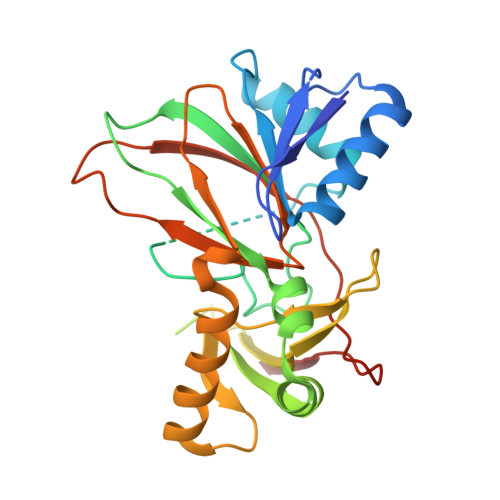
    Rv3406 evolved from the ubiquitous taurine-catabolising enzyme TauD and functions as a sulfur-scavenging protein in Mycobacterium tuberculosis . Structural and biochemical analyses reveal specific changes that shape its chemical environment for ligand interaction and explain its broad substrate range. These findings show how amino acid substitutions redefine protein function and drive adaptation to the unique metabolic context of Mycobacteria.


  • Organizational Affiliation
    • School of Chemical Sciences, The University of Auckland, Auckland, 1142, New Zealand.

Macromolecule Content 

  • Total Structure Weight: 132.55 kDa 
  • Atom Count: 7,292 
  • Modeled Residue Count: 943 
  • Deposited Residue Count: 1,192 
  • Unique protein chains: 1

Macromolecules

Find similar proteins by:|  3D Structure
Entity ID: 1
MoleculeChains  Sequence LengthOrganismDetailsImage
Alpha-ketoglutarate-dependent sulfate ester dioxygenase
A, B, C, D
298Mycobacterium tuberculosisMutation(s): 0 
Gene Names: Rv3406MTCY78.22c
EC: 1.14.11 (PDB Primary Data), 1.14.11.77 (UniProt)
UniProt
Find proteins for P9WKZ1 (Mycobacterium tuberculosis (strain ATCC 25618 / H37Rv))
Explore P9WKZ1 
Go to UniProtKB:  P9WKZ1
Entity Groups
Sequence Clusters30% Identity50% Identity70% Identity90% Identity95% Identity100% Identity
UniProt GroupP9WKZ1
Sequence Annotations
Expand
Reference Sequence

Small Molecules

Ligands 3 Unique
IDChains Name / Formula / InChI Key2D Diagram3D Interactions
OGA
(Subject of Investigation/LOI)

Query on OGA


Download:Ideal Coordinates CCD File
E [auth A],
G [auth B],
I [auth C],
K [auth D]
N-OXALYLGLYCINE
C4 H5 N O5
BIMZLRFONYSTPT-UHFFFAOYSA-N
TRS

Query on TRS


Download:Ideal Coordinates CCD File
L [auth D]2-AMINO-2-HYDROXYMETHYL-PROPANE-1,3-DIOL
C4 H12 N O3
LENZDBCJOHFCAS-UHFFFAOYSA-O
NI

Query on NI


Download:Ideal Coordinates CCD File
F [auth A],
H [auth B],
J [auth C],
M [auth D]
NICKEL (II) ION
Ni
VEQPNABPJHWNSG-UHFFFAOYSA-N

Experimental Data & Validation

Experimental Data

  • Method: X-RAY DIFFRACTION
  • Resolution: 2.64 Å
  • R-Value Free:  0.299 (Depositor), 0.314 (DCC) 
  • R-Value Work:  0.250 (Depositor), 0.267 (DCC) 
Space Group: P 21 21 21
Unit Cell:
Length ( Å )Angle ( ˚ )
a = 65.535α = 90
b = 128.336β = 90
c = 138.693γ = 90
Software Package:
Software NamePurpose
REFMACrefinement
XDSdata reduction
Aimlessdata scaling
PHASERphasing

Structure Validation

View Full Validation Report


Ligand Structure Quality Assessment 


Entry History 

& Funding Information

Deposition Data


Funding OrganizationLocationGrant Number
Not fundedNew Zealand--

Revision History  (Full details and data files)

  • Version 1.0: 2023-10-25
    Type: Initial release
  • Version 1.1: 2024-11-20
    Changes: Structure summary
  • Version 1.2: 2026-03-04
    Changes: Refinement description
  • Version 1.3: 2026-03-18
    Changes: Database references