


Structural diversity of bile salt hydrolases reveals rationale for substrate selectivity
Walker, M.E., Lim, L., Redinbo, M.R.To be published.
Experimental Data Snapshot
Starting Model: experimental
View more details
Entity ID: 1 | |||||
|---|---|---|---|---|---|
| Molecule | Chains | Sequence Length | Organism | Details | Image |
| Conjugated bile acid hydrolase | 317 | Bifidobacterium longum | Mutation(s): 0 Gene Names: bsh EC: 3.5.1 (PDB Primary Data), 3.5.1.74 (PDB Primary Data), 3.5.1.24 (PDB Primary Data), 2.3.1 (UniProt) | 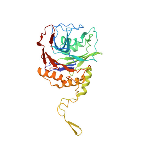 | |
UniProt | |||||
Entity Groups | |||||
| Sequence Clusters | 30% Identity50% Identity70% Identity90% Identity95% Identity100% Identity | ||||
| UniProt Group | Q9KK62 | ||||
Sequence AnnotationsExpand | |||||
Reference Sequence | |||||
| Ligands 1 Unique | |||||
|---|---|---|---|---|---|
| ID | Chains | Name / Formula / InChI Key | 2D Diagram | 3D Interactions | |
| WSR (Subject of Investigation/LOI) Download:Ideal Coordinates CCD File | I [auth A] J [auth B] K [auth C] L [auth D] M [auth E] | (1R,3aS,3bR,5aR,7R,9aS,9bS,11aR)-1-[(2R)-6-fluoro-5-oxohexan-2-yl]-9a,11a-dimethylhexadecahydro-1H-cyclopenta[a]phenanthren-7-yl hydrogen sulfate (non-preferred name) C25 H41 F O5 S MVPISWRWXKQVBV-CIQXVCGBSA-N |  | ||
| Length ( Å ) | Angle ( ˚ ) |
|---|---|
| a = 93.072 | α = 90 |
| b = 166.801 | β = 116.6 |
| c = 103.077 | γ = 90 |
| Software Name | Purpose |
|---|---|
| PHENIX | refinement |
| PHENIX | refinement |
| XDS | data reduction |
| XDS | data scaling |
| PHASER | phasing |
| Funding Organization | Location | Grant Number |
|---|---|---|
| National Institutes of Health/National Institute of General Medical Sciences (NIH/NIGMS) | United States | GM135218 |


