8EML | pdb_00008eml

Crystal Structure of Gsx2 Homeodomain in Complex with DNA


Experimental Data Snapshot

  • Method: X-RAY DIFFRACTION
  • Resolution: 2.21 Å
  • R-Value Free: 
    0.265 (Depositor), 0.265 (DCC) 
  • R-Value Work: 
    0.220 (Depositor), 0.220 (DCC) 
  • R-Value Observed: 
    0.225 (Depositor) 

Starting Model: experimental
View more details

wwPDB Validation 3D Report Full Report

Validation slider image for 8EML

This is version 1.2 of the entry. See complete history

Literature

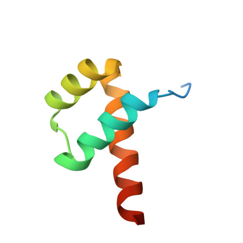
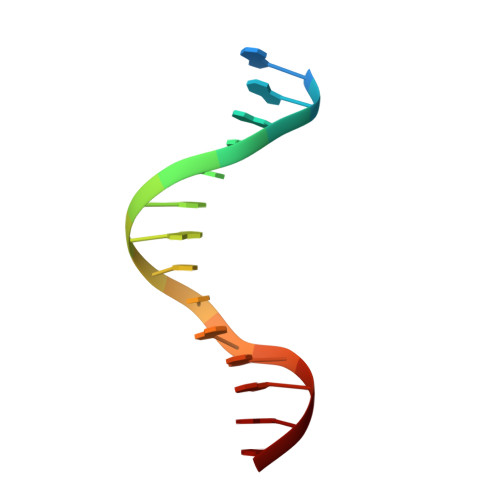
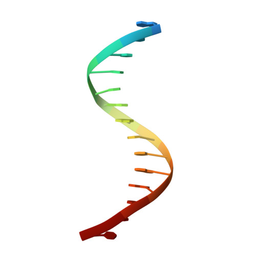
Cooperative Gsx2-DNA binding requires DNA bending and a novel Gsx2 homeodomain interface.

Webb, J.A.Farrow, E.Cain, B.Yuan, Z.Yarawsky, A.E.Schoch, E.Gagliani, E.K.Herr, A.B.Gebelein, B.Kovall, R.A.

(2024) Nucleic Acids Res 52: 7987-8002

  • DOI: https://doi.org/10.1093/nar/gkae522
  • Primary Citation Related Structures: 
    8EML

  • PubMed Abstract: 

    The conserved Gsx homeodomain (HD) transcription factors specify neural cell fates in animals from flies to mammals. Like many HD proteins, Gsx factors bind A/T-rich DNA sequences prompting the following question: How do HD factors that bind similar DNA sequences in vitro regulate specific target genes in vivo? Prior studies revealed that Gsx factors bind DNA both as a monomer on individual A/T-rich sites and as a cooperative homodimer to two sites spaced precisely 7 bp apart. However, the mechanistic basis for Gsx-DNA binding and cooperativity is poorly understood. Here, we used biochemical, biophysical, structural and modeling approaches to (i) show that Gsx factors are monomers in solution and require DNA for cooperative complex formation, (ii) define the affinity and thermodynamic binding parameters of Gsx2/DNA interactions, (iii) solve a high-resolution monomer/DNA structure that reveals that Gsx2 induces a 20° bend in DNA, (iv) identify a Gsx2 protein-protein interface required for cooperative DNA binding and (v) determine that flexible spacer DNA sequences enhance Gsx2 cooperativity on dimer sites. Altogether, our results provide a mechanistic basis for understanding the protein and DNA structural determinants that underlie cooperative DNA binding by Gsx factors.


  • Organizational Affiliation
    • Department of Molecular and Cellular Biosciences, University of Cincinnati College of Medicine, Cincinnati, OH 45267, USA.

Macromolecule Content 

  • Total Structure Weight: 34.27 kDa 
  • Atom Count: 2,123 
  • Modeled Residue Count: 171 
  • Deposited Residue Count: 194 
  • Unique protein chains: 1
  • Unique nucleic acid chains: 2

Macromolecules


Find similar proteins by:|  3D Structure
Entity ID: 1
MoleculeChains  Sequence LengthOrganismDetailsImage
GS homeobox 2A [auth B],
D [auth A]
67Mus musculusMutation(s): 0 
Gene Names: Gsx2Gsh-2Gsh2
UniProt
Find proteins for P31316 (Mus musculus)
Explore P31316 
Go to UniProtKB:  P31316
Entity Groups
Sequence Clusters30% Identity50% Identity70% Identity90% Identity95% Identity100% Identity
UniProt GroupP31316
Sequence Annotations
Expand
Reference Sequence
Find similar nucleic acids by:  Sequence
Entity ID: 2
MoleculeChains LengthOrganismImage
DNA (5'-D(P*GP*AP*GP*CP*TP*AP*AP*TP*TP*AP*AP*AP*GP*C)-3')B [auth E],
E [auth C]
15Mus musculus
Sequence Annotations
Expand
Reference Sequence
Find similar nucleic acids by:  Sequence
Entity ID: 3
MoleculeChains LengthOrganismImage
DNA (5'-D(P*GP*CP*TP*TP*TP*AP*AP*TP*TP*AP*GP*CP*TP*C)-3')C [auth F],
F [auth D]
15Mus musculus
Sequence Annotations
Expand
Reference Sequence

Small Molecules

Ligands 1 Unique
IDChains Name / Formula / InChI Key2D Diagram3D Interactions
PGE

Query on PGE


Download:Ideal Coordinates CCD File
G [auth A]TRIETHYLENE GLYCOL
C6 H14 O4
ZIBGPFATKBEMQZ-UHFFFAOYSA-N

Experimental Data & Validation

Experimental Data

  • Method: X-RAY DIFFRACTION
  • Resolution: 2.21 Å
  • R-Value Free:  0.265 (Depositor), 0.265 (DCC) 
  • R-Value Work:  0.220 (Depositor), 0.220 (DCC) 
  • R-Value Observed: 0.225 (Depositor) 
Space Group: P 1 21 1
Unit Cell:
Length ( Å )Angle ( ˚ )
a = 37.7α = 90
b = 37.65β = 93.99
c = 107.869γ = 90
Software Package:
Software NamePurpose
PHENIXrefinement
MOSFLMdata reduction
Aimlessdata scaling
PHASERphasing

Structure Validation

View Full Validation Report


Entry History 

& Funding Information

Deposition Data


Funding OrganizationLocationGrant Number
National Institutes of Health/National Institute of General Medical Sciences (NIH/NIGMS)United StatesGM079428

Revision History  (Full details and data files)

  • Version 1.0: 2023-10-18
    Type: Initial release
  • Version 1.1: 2024-10-30
    Changes: Database references, Structure summary
  • Version 1.2: 2026-03-04
    Changes: Refinement description