8EM4 | pdb_00008em4

Cryo-EM structure of LRP2 at pH 7.5


Experimental Data Snapshot

  • Method: ELECTRON MICROSCOPY
  • Resolution: 2.83 Å
  • Aggregation State: PARTICLE 
  • Reconstruction Method: SINGLE PARTICLE 

wwPDB Validation 3D Report Full Report

Validation slider image for 8EM4

This is version 1.3 of the entry. See complete history

Literature

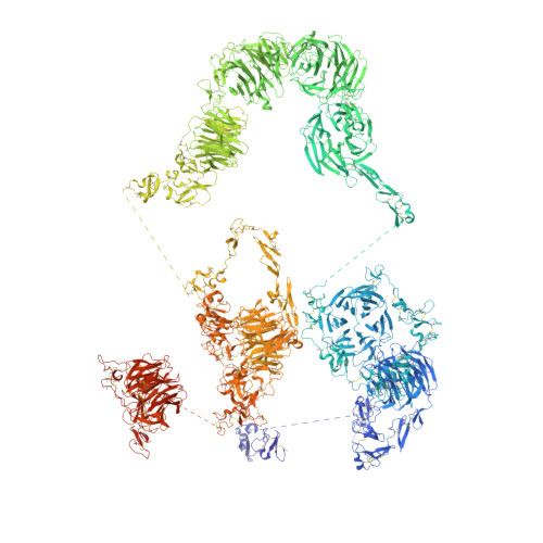
Structures of LRP2 reveal a molecular machine for endocytosis.

Beenken, A.Cerutti, G.Brasch, J.Guo, Y.Sheng, Z.Erdjument-Bromage, H.Aziz, Z.Robbins-Juarez, S.Y.Chavez, E.Y.Ahlsen, G.Katsamba, P.S.Neubert, T.A.Fitzpatrick, A.W.P.Barasch, J.Shapiro, L.

(2023) Cell 186: 821

  • DOI: https://doi.org/10.1016/j.cell.2023.01.016
  • Primary Citation Related Structures: 
    8EM4, 8EM7

  • PubMed Abstract: 

    The low-density lipoprotein (LDL) receptor-related protein 2 (LRP2 or megalin) is representative of the phylogenetically conserved subfamily of giant LDL receptor-related proteins, which function in endocytosis and are implicated in diseases of the kidney and brain. Here, we report high-resolution cryoelectron microscopy structures of LRP2 isolated from mouse kidney, at extracellular and endosomal pH. The structures reveal LRP2 to be a molecular machine that adopts a conformation for ligand binding at the cell surface and for ligand shedding in the endosome. LRP2 forms a homodimer, the conformational transformation of which is governed by pH-sensitive sites at both homodimer and intra-protomer interfaces. A subset of LRP2 deleterious missense variants in humans appears to impair homodimer assembly. These observations lay the foundation for further understanding the function and mechanism of LDL receptors and implicate homodimerization as a conserved feature of the LRP receptor subfamily.


  • Organizational Affiliation
    • Division of Nephrology, Department of Medicine, Columbia University Vagelos College of Physicians and Surgeons, New York, NY 10032, USA.

Macromolecule Content 

  • Total Structure Weight: 1,063.62 kDa 
  • Atom Count: 59,410 
  • Modeled Residue Count: 7,636 
  • Deposited Residue Count: 9,320 
  • Unique protein chains: 1

Macromolecules

Find similar proteins by:|  3D Structure
Entity ID: 1
MoleculeChains  Sequence LengthOrganismDetailsImage
Low-density lipoprotein receptor-related protein 2
A, B
4,660Mus musculusMutation(s): 0 
UniProt
Find proteins for A2ARV4 (Mus musculus)
Explore A2ARV4 
Go to UniProtKB:  A2ARV4
Entity Groups
Sequence Clusters30% Identity50% Identity70% Identity90% Identity95% Identity100% Identity
UniProt GroupA2ARV4
Glycosylation
Glycosylation Sites: 43Go to GlyGen: A2ARV4-1
Sequence Annotations
Expand
Reference Sequence

Oligosaccharides

Help  
Entity ID: 2
MoleculeChains Length2D Diagram GlycosylationD Interactions
2-acetamido-2-deoxy-beta-D-glucopyranose-(1-4)-2-acetamido-2-deoxy-beta-D-glucopyranose
C, D
2N-Glycosylation
Glycosylation Resources
GlyTouCan: G42666HT
GlyCosmos: G42666HT
GlyGen: G42666HT

Small Molecules

Ligands 3 Unique
IDChains Name / Formula / InChI Key2D Diagram3D Interactions
NAG
(Subject of Investigation/LOI)

Query on NAG


Download:Ideal Coordinates CCD File
AA [auth A]
AD [auth B]
BA [auth A]
BE [auth B]
CA [auth A]
AA [auth A],
AD [auth B],
BA [auth A],
BE [auth B],
CA [auth A],
CE [auth B],
DA [auth A],
DD [auth B],
EA [auth A],
ED [auth B],
EE [auth B],
F [auth A],
FA [auth A],
FD [auth B],
G [auth A],
GA [auth A],
GD [auth B],
H [auth A],
I [auth A],
ID [auth B],
IE [auth B],
J [auth A],
JD [auth B],
K [auth A],
KF [auth B],
L [auth A],
M [auth A],
MC [auth A],
MF [auth B],
N [auth A],
NC [auth B],
ND [auth B],
O [auth A],
OD [auth B],
P [auth A],
Q [auth A],
QD [auth B],
R [auth A],
RD [auth B],
S [auth A],
SC [auth B],
T [auth A],
TD [auth B],
TF [auth B],
U [auth A],
UC [auth B],
UD [auth B],
V [auth A],
VC [auth B],
VD [auth B],
W [auth A],
WC [auth B],
WD [auth B],
X [auth A],
XC [auth B],
Y [auth A],
Z [auth A],
ZD [auth B]
2-acetamido-2-deoxy-beta-D-glucopyranose
C8 H15 N O6
OVRNDRQMDRJTHS-FMDGEEDCSA-N
NGA
(Subject of Investigation/LOI)

Query on NGA


Download:Ideal Coordinates CCD File
AE [auth B]
E [auth A]
EF [auth B]
FE [auth B]
HA [auth A]
AE [auth B],
E [auth A],
EF [auth B],
FE [auth B],
HA [auth A],
HD [auth B],
IA [auth A],
JA [auth A],
KA [auth A],
LA [auth A],
LD [auth B],
MA [auth A],
MD [auth B],
NA [auth A],
OA [auth A],
OC [auth B],
OF [auth B],
PA [auth A],
PD [auth B],
QA [auth A],
QF [auth B],
RA [auth A],
RC [auth B],
RF [auth B],
SA [auth A],
SF [auth B],
TA [auth A],
UA [auth A],
VA [auth A],
VF [auth B],
YC [auth B],
YD [auth B]
2-acetamido-2-deoxy-beta-D-galactopyranose
C8 H15 N O6
OVRNDRQMDRJTHS-JAJWTYFOSA-N
CA
(Subject of Investigation/LOI)

Query on CA


Download:Ideal Coordinates CCD File
AB [auth A]
AC [auth A]
AF [auth B]
BB [auth A]
BC [auth A]
AB [auth A],
AC [auth A],
AF [auth B],
BB [auth A],
BC [auth A],
BD [auth B],
BF [auth B],
CB [auth A],
CC [auth A],
CD [auth B],
CF [auth B],
DB [auth A],
DC [auth A],
DE [auth B],
DF [auth B],
EB [auth A],
EC [auth A],
FB [auth A],
FC [auth A],
FF [auth B],
GB [auth A],
GC [auth A],
GE [auth B],
GF [auth B],
HB [auth A],
HC [auth A],
HE [auth B],
HF [auth B],
IB [auth A],
IC [auth A],
IF [auth B],
JB [auth A],
JC [auth A],
JE [auth B],
JF [auth B],
KB [auth A],
KC [auth A],
KD [auth B],
KE [auth B],
LB [auth A],
LC [auth A],
LE [auth B],
LF [auth B],
MB [auth A],
ME [auth B],
NB [auth A],
NE [auth B],
NF [auth B],
OB [auth A],
OE [auth B],
PB [auth A],
PC [auth B],
PE [auth B],
PF [auth B],
QB [auth A],
QC [auth B],
QE [auth B],
RB [auth A],
RE [auth B],
SB [auth A],
SD [auth B],
SE [auth B],
TB [auth A],
TC [auth B],
TE [auth B],
UB [auth A],
UE [auth B],
UF [auth B],
VB [auth A],
VE [auth B],
WA [auth A],
WB [auth A],
WE [auth B],
XA [auth A],
XB [auth A],
XD [auth B],
XE [auth B],
YA [auth A],
YB [auth A],
YE [auth B],
ZA [auth A],
ZB [auth A],
ZC [auth B],
ZE [auth B]
CALCIUM ION
Ca
BHPQYMZQTOCNFJ-UHFFFAOYSA-N

Experimental Data & Validation

Experimental Data

  • Method: ELECTRON MICROSCOPY
  • Resolution: 2.83 Å
  • Aggregation State: PARTICLE 
  • Reconstruction Method: SINGLE PARTICLE 
EM Software:
TaskSoftware PackageVersion
RECONSTRUCTIONcryoSPARC

Structure Validation

View Full Validation Report


Entry History 

& Funding Information

Deposition Data


Funding OrganizationLocationGrant Number
National Institutes of Health/National Institute of Diabetes and Digestive and Kidney Disease (NIH/NIDDK)United StatesDK124667

Revision History  (Full details and data files)

  • Version 1.0: 2023-02-08
    Type: Initial release
  • Version 1.1: 2023-02-22
    Changes: Database references, Structure summary
  • Version 1.2: 2023-03-08
    Changes: Database references
  • Version 1.3: 2024-11-13
    Changes: Data collection, Structure summary