8EJN | pdb_00008ejn

Structure of dehaloperoxidase A in complex with 2,4-dichlorophenol


Experimental Data Snapshot

  • Method: X-RAY DIFFRACTION
  • Resolution: 1.48 Å
  • R-Value Free: 
    0.211 (Depositor), 0.211 (DCC) 
  • R-Value Work: 
    0.160 (Depositor), 0.160 (DCC) 

Starting Model: experimental
View more details

wwPDB Validation 3D Report Full Report

Validation slider image for 8EJN

Ligand Structure Quality Assessment 


This is version 1.2 of the entry. See complete history

Literature

Comparative study of the binding and activation of 2,4-dichlorophenol by dehaloperoxidase A and B.

Aktar, M.S.de Serrano, V.Ghiladi, R.Franzen, S.

(2023) J Inorg Biochem 247: 112332-112332

  • DOI: https://doi.org/10.1016/j.jinorgbio.2023.112332
  • Primary Citation Related Structures: 
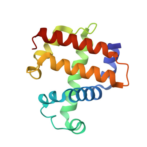
    8EJN

  • PubMed Abstract: 

    The dehaloperoxidase-hemoglobin (DHP), first isolated from the coelom of a marine terebellid polychaete, Amphitrite ornata, is an example of a multi-functional heme enzyme. Long known for its reversible oxygen (O 2 ) binding, further studies have established DHP activity as a peroxidase, oxidase, oxygenase, and peroxygenase. The specific reactivity depends on substrate binding at various internal and external binding sites. This study focuses on comparison of the binding and reactivity of the substrate 2,4-dichlorophenol (DCP) in the isoforms DHPA and B. There is strong interest in the degradation of DCP because of its wide use in the chemical industry, presence in waste streams, and particular reactivity to form dioxins, some of the most toxic compounds known. The catalytic efficiency is 3.5 times higher for DCP oxidation in DHPB than DHPA by a peroxidase mechanism. However, DHPA and B both show self-inhibition even at modest concentrations of DCP. This phenomenon is analogous to the self-inhibition of 2,4,6-trichlorophenol (TCP) at higher concentration. The activation energies of the electron transfer steps in DCP in DHPA and DHPB are 19.3 ± 2.5 and 24.3 ± 3.2 kJ/mol, respectively, compared to 37.2 ± 6.5 kJ/mol in horseradish peroxidase (HRP), which may be a result of the more facile electron transfer of an internally bound substrate in DHPA. The x-ray crystal structure of DHPA bound with DCP determined at 1.48 Å resolution, shows tight substrate binding inside the heme pocket of DHPA (PDB 8EJN). This research contributes to the studies of DHP as a naturally occurring bioremediation enzyme capable of oxidizing a wide range of environmental pollutants.


  • Organizational Affiliation
    • Department of Chemistry, North Carolina State University, Raleigh, NC 27695, United States of America.

Macromolecule Content 

  • Total Structure Weight: 33.28 kDa 
  • Atom Count: 2,775 
  • Modeled Residue Count: 274 
  • Deposited Residue Count: 274 
  • Unique protein chains: 1

Macromolecules

Find similar proteins by:|  3D Structure
Entity ID: 1
MoleculeChains  Sequence LengthOrganismDetailsImage
Dehaloperoxidase A
A, B
137Amphitrite ornataMutation(s): 0 
UniProt
Find proteins for Q9NAV8 (Amphitrite ornata)
Explore Q9NAV8 
Go to UniProtKB:  Q9NAV8
Entity Groups
Sequence Clusters30% Identity50% Identity70% Identity90% Identity95% Identity100% Identity
UniProt GroupQ9NAV8
Sequence Annotations
Expand
Reference Sequence

Small Molecules

Ligands 5 Unique
IDChains Name / Formula / InChI Key2D Diagram3D Interactions
HEM

Query on HEM


Download:Ideal Coordinates CCD File
C [auth A],
G [auth B]
PROTOPORPHYRIN IX CONTAINING FE
C34 H32 Fe N4 O4
KABFMIBPWCXCRK-RGGAHWMASA-L
5JC

Query on 5JC


Download:Ideal Coordinates CCD File
D [auth A],
H [auth B]
2,4-dichlorophenol
C6 H4 Cl2 O
HFZWRUODUSTPEG-UHFFFAOYSA-N
SO4

Query on SO4


Download:Ideal Coordinates CCD File
F [auth A],
M [auth B]
SULFATE ION
O4 S
QAOWNCQODCNURD-UHFFFAOYSA-L
GOL

Query on GOL


Download:Ideal Coordinates CCD File
E [auth A],
I [auth B],
J [auth B]
GLYCEROL
C3 H8 O3
PEDCQBHIVMGVHV-UHFFFAOYSA-N
DMS

Query on DMS


Download:Ideal Coordinates CCD File
K [auth B],
L [auth B]
DIMETHYL SULFOXIDE
C2 H6 O S
IAZDPXIOMUYVGZ-UHFFFAOYSA-N

Experimental Data & Validation

Experimental Data

  • Method: X-RAY DIFFRACTION
  • Resolution: 1.48 Å
  • R-Value Free:  0.211 (Depositor), 0.211 (DCC) 
  • R-Value Work:  0.160 (Depositor), 0.160 (DCC) 
Space Group: P 21 21 21
Unit Cell:
Length ( Å )Angle ( ˚ )
a = 57.881α = 90
b = 67.323β = 90
c = 69.199γ = 90
Software Package:
Software NamePurpose
REFMACrefinement
HKL-2000data reduction
HKL-2000data scaling
PHASERphasing

Structure Validation

View Full Validation Report


Ligand Structure Quality Assessment 


Entry History 

& Funding Information

Deposition Data


Funding OrganizationLocationGrant Number
National Natural Science Foundation of China (NSFC)China--

Revision History  (Full details and data files)

  • Version 1.0: 2023-08-23
    Type: Initial release
  • Version 1.1: 2023-08-30
    Changes: Derived calculations, Structure summary
  • Version 1.2: 2026-03-04
    Changes: Refinement description, Structure summary