8EFX

Structure of OtDUB DUB Domain disulfide crosslinked with Ubiquitin


Experimental Data Snapshot

  • Method: X-RAY DIFFRACTION
  • Resolution: 1.85 Å
  • R-Value Free: 0.229 
  • R-Value Work: 0.198 
  • R-Value Observed: 0.200 

Starting Models: experimental
View more details

wwPDB Validation   3D Report Full Report


This is version 1.2 of the entry. See complete history


Literature

Cocrystallization of ubiquitin-deubiquitinase complexes through disulfide linkage.

Negron Teron, K.I.Das, C.

(2023) Acta Crystallogr D Struct Biol 79: 1044-1055

  • DOI: https://doi.org/10.1107/S2059798323008501
  • Primary Citation of Related Structures:  
    8EFW, 8EFX

  • PubMed Abstract: 

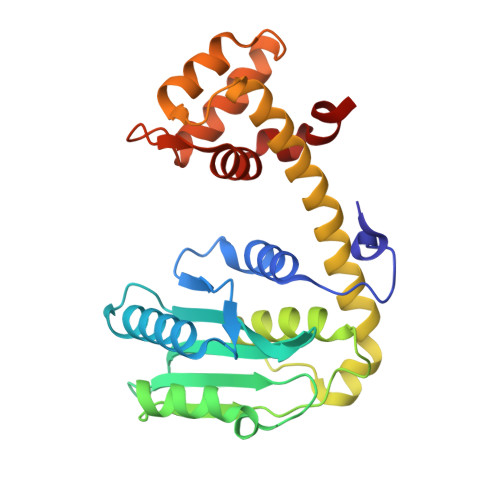
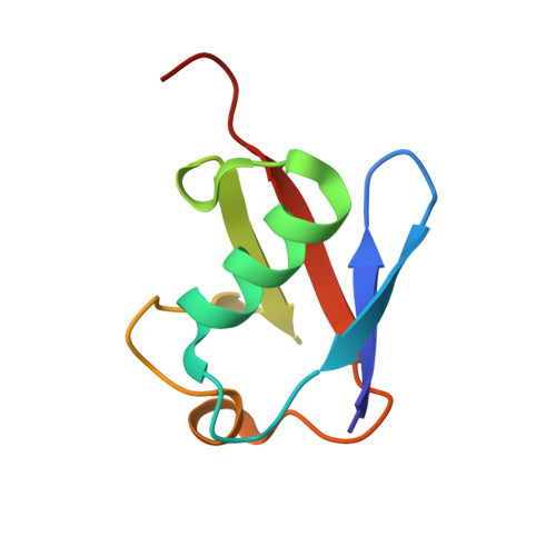
    Structural characterization of the recognition of ubiquitin (Ub) by deubiquitinases (DUBs) has largely relied on covalent complexation of the DUB through its catalytic cysteine with a Ub C-terminal electrophile. The Ub electrophiles are accessed through intein chemistry in conjunction with chemical synthesis. Here, it was asked whether DUB-Ub covalent complexes could instead be accessed by simpler disulfide chemistry using a Ub cysteine mutant in which the last glycine has been replaced with a cysteine. The Ub cysteine mutant displayed a wide variability in disulfide formation across a panel of eukaryotic and prokaryotic DUBs, with some showing no detectable reaction while others robustly produced a disulfide complex. Using this approach, two disulfide-linked ubiquitin-bound complexes were crystallized, one involving the Legionella pneumophila effector SdeA DUB and the other involving the Orientia effector OtDUB. These DUBs had previously been crystallized in Ub-bound forms using the C-terminal electrophile strategy and noncovalent complexation, respectively. While the disulfide-linked SdeA DUB-Ub complex crystallized as expected, in the OtDUB complex the disulfide bond to the Ub mutant involved a cysteine that differed from the catalytic cysteine. Disulfide formation with the SdeA DUB catalytic cysteine was accompanied by local distortion of the helix carrying the active-site cysteine, whereas OtDUB reacted with the Ub mutant using a surface-exposed cysteine.


  • Organizational Affiliation

    Department of Chemistry, Purdue University, 560 Oval Drive, West Lafayette, IN 47907, USA.


Macromolecules
Find similar proteins by:  (by identity cutoff)  |  3D Structure
Entity ID: 1
MoleculeChains Sequence LengthOrganismDetailsImage
OtDUB259Orientia tsutsugamushiMutation(s): 0 
Gene Names: KATO_02133
UniProt
Find proteins for A0A2U3QWA1 (Orientia tsutsugamushi)
Explore A0A2U3QWA1 
Go to UniProtKB:  A0A2U3QWA1
Entity Groups  
Sequence Clusters30% Identity50% Identity70% Identity90% Identity95% Identity100% Identity
UniProt GroupA0A2U3QWA1
Sequence Annotations
Expand
  • Reference Sequence
Find similar proteins by:  (by identity cutoff)  |  3D Structure
Entity ID: 2
MoleculeChains Sequence LengthOrganismDetailsImage
Ubiquitin76Homo sapiensMutation(s): 1 
Gene Names: UBB
UniProt & NIH Common Fund Data Resources
Find proteins for P0CG47 (Homo sapiens)
Explore P0CG47 
Go to UniProtKB:  P0CG47
GTEx:  ENSG00000170315 
Entity Groups  
Sequence Clusters30% Identity50% Identity70% Identity90% Identity95% Identity100% Identity
UniProt GroupP0CG47
Sequence Annotations
Expand
  • Reference Sequence
Experimental Data & Validation

Experimental Data

  • Method: X-RAY DIFFRACTION
  • Resolution: 1.85 Å
  • R-Value Free: 0.229 
  • R-Value Work: 0.198 
  • R-Value Observed: 0.200 
  • Space Group: P 2 21 21
Unit Cell:
Length ( Å )Angle ( ˚ )
a = 42.399α = 90
b = 83.008β = 90
c = 99.354γ = 90
Software Package:
Software NamePurpose
PDB_EXTRACTdata extraction
PHENIXrefinement
HKL-3000data reduction
HKL-3000data scaling
HKL-3000phasing

Structure Validation

View Full Validation Report


Entry History & Funding Information

Deposition Data


Funding OrganizationLocationGrant Number
National Institutes of Health/National Institute of General Medical Sciences (NIH/NIGMS)United States5R01GM126296-02

Revision History  (Full details and data files)

  • Version 1.0: 2023-09-20
    Type: Initial release
  • Version 1.1: 2023-11-08
    Changes: Database references
  • Version 1.2: 2024-05-01
    Changes: Database references