


The crystal structure of I38T mutant PA endonuclease (2009/H1N1/CALIFORNIA) in complex with compound SJ000986319
Cuypers, M.G., Slavish, J.P., Rankovic, Z., White, S.W.To be published.
Experimental Data Snapshot
Starting Model: experimental
View more details
Entity ID: 1 | |||||
|---|---|---|---|---|---|
| Molecule | Chains | Sequence Length | Organism | Details | Image |
| Polymerase acidic protein | 197 | Influenza A virus (A/Luxembourg/43/2009(H1N1)) | Mutation(s): 1 Gene Names: PA | 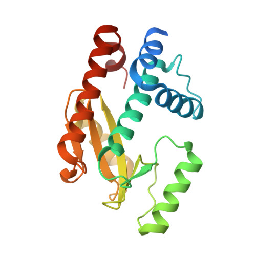 | |
UniProt | |||||
Entity Groups | |||||
| Sequence Clusters | 30% Identity50% Identity70% Identity90% Identity95% Identity100% Identity | ||||
| UniProt Group | C6H0Y9 | ||||
Sequence AnnotationsExpand | |||||
Reference Sequence | |||||
| Ligands 4 Unique | |||||
|---|---|---|---|---|---|
| ID | Chains | Name / Formula / InChI Key | 2D Diagram | 3D Interactions | |
| QQ4 Download:Ideal Coordinates CCD File | C [auth A] | Hexa Vinylpyrrolidone K15 C36 H56 N6 O6 OFPQNVWJHZVWCZ-CMPUJJQDSA-N |  | ||
| WFW (Subject of Investigation/LOI) Download:Ideal Coordinates CCD File | B [auth A] | benzyl [2-(6-{[2-(4-amino-7H-pyrrolo[2,3-d]pyrimidin-7-yl)ethyl]carbamoyl}-5-hydroxy-4-oxo-1,4-dihydropyrimidin-2-yl)propan-2-yl]carbamate C24 H26 N8 O5 NCRFYYQIIFPHLD-UHFFFAOYSA-N |  | ||
| SO4 Download:Ideal Coordinates CCD File | F [auth A] | SULFATE ION O4 S QAOWNCQODCNURD-UHFFFAOYSA-L |  | ||
| MN Download:Ideal Coordinates CCD File | D [auth A], E [auth A] | MANGANESE (II) ION Mn WAEMQWOKJMHJLA-UHFFFAOYSA-N |  | ||
| Length ( Å ) | Angle ( ˚ ) |
|---|---|
| a = 89.688 | α = 90 |
| b = 89.688 | β = 90 |
| c = 132.251 | γ = 90 |
| Software Name | Purpose |
|---|---|
| PHENIX | refinement |
| XDS | data reduction |
| Aimless | data scaling |
| MOLREP | phasing |
| Funding Organization | Location | Grant Number |
|---|---|---|
| National Institutes of Health/National Cancer Institute (NIH/NCI) | United States | -- |


