8ECD | pdb_00008ecd

E. coli L-asparaginase II mutant (V27T) in complex with L-Asp


Experimental Data Snapshot

  • Method: X-RAY DIFFRACTION
  • Resolution: 1.62 Å
  • R-Value Free: 
    0.164 (Depositor), 0.170 (DCC) 
  • R-Value Work: 
    0.127 (Depositor), 0.133 (DCC) 
  • R-Value Observed: 
    0.127 (Depositor) 

Starting Model: experimental
View more details

wwPDB Validation 3D Report Full Report

Validation slider image for 8ECD

Ligand Structure Quality Assessment 


This is version 1.3 of the entry. See complete history

Literature

The E. coli L-asparaginase V27T mutant: structural and functional characterization and comparison with theoretical predictions.

Strzelczyk, P.Zhang, D.Wlodawer, A.Lubkowski, J.

(2022) FEBS Lett 596: 3060-3068

  • DOI: https://doi.org/10.1002/1873-3468.14526
  • Primary Citation Related Structures: 
    8ECD, 8ECE

  • PubMed Abstract: 

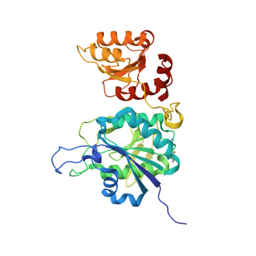
    Bacterial L-asparaginases have been used for over 40 years as anticancer drugs. Ardalan et al. (Medical Hypotheses 112, 7-17, 2018) proposed that the V27T mutant of Escherichia coli type II L-asparaginase, EcAII(V27T), should display altered biophysical and catalytic properties compared to the wild-type enzyme, EcAII(wt), rendering it more favourable as a pharmaceutical. They postulated that EcAII(V27T) would exhibit reduced glutaminolytic activity and be more stable compared to EcAII(wt). Their postulates, however, were purely theoretical. Here, we characterized experimentally selected properties of EcAII(V27T). We found asparaginolytic activity of this mutant unchanged, whereas its glutaminolytic activity was fourfold lower compared with EcAII(wt). We did not observe significant differences in stabilities of EcAII(wt) and EcAII(V27T). Crystal structures of the complexes with L-Asp and L-Glu showed considerable differences in binding modes of both substrates.


  • Organizational Affiliation
    • Center for Structural Biology, Center for Cancer Research, National Cancer Institute, Frederick, MD, USA.

Macromolecule Content 

  • Total Structure Weight: 145.12 kDa 
  • Atom Count: 11,603 
  • Modeled Residue Count: 1,309 
  • Deposited Residue Count: 1,336 
  • Unique protein chains: 1

Macromolecules

Find similar proteins by:|  3D Structure
Entity ID: 1
MoleculeChains  Sequence LengthOrganismDetailsImage
L-asparaginase 2
A, B, C, D
334Escherichia coli K-12Mutation(s): 1 
Gene Names: ansBb2957JW2924
EC: 3.5.1.1
UniProt
Find proteins for P00805 (Escherichia coli (strain K12))
Explore P00805 
Go to UniProtKB:  P00805
Entity Groups
Sequence Clusters30% Identity50% Identity70% Identity90% Identity95% Identity100% Identity
UniProt GroupP00805
Sequence Annotations
Expand
Reference Sequence

Small Molecules

Ligands 4 Unique
IDChains Name / Formula / InChI Key2D Diagram3D Interactions
CIT

Query on CIT


Download:Ideal Coordinates CCD File
U [auth C],
V [auth D]
CITRIC ACID
C6 H8 O7
KRKNYBCHXYNGOX-UHFFFAOYSA-N
ASP
(Subject of Investigation/LOI)

Query on ASP


Download:Ideal Coordinates CCD File
E [auth A],
I [auth B],
M [auth C],
W [auth D]
ASPARTIC ACID
C4 H7 N O4
CKLJMWTZIZZHCS-REOHCLBHSA-N
GOL

Query on GOL


Download:Ideal Coordinates CCD File
AA [auth D]
CA [auth D]
F [auth A]
J [auth B]
K [auth B]
AA [auth D],
CA [auth D],
F [auth A],
J [auth B],
K [auth B],
N [auth C],
X [auth D]
GLYCEROL
C3 H8 O3
PEDCQBHIVMGVHV-UHFFFAOYSA-N
EDO

Query on EDO


Download:Ideal Coordinates CCD File
BA [auth D]
G [auth A]
H [auth A]
L [auth B]
O [auth C]
BA [auth D],
G [auth A],
H [auth A],
L [auth B],
O [auth C],
P [auth C],
Q [auth C],
R [auth C],
S [auth C],
T [auth C],
Y [auth D],
Z [auth D]
1,2-ETHANEDIOL
C2 H6 O2
LYCAIKOWRPUZTN-UHFFFAOYSA-N

Experimental Data & Validation

Experimental Data

  • Method: X-RAY DIFFRACTION
  • Resolution: 1.62 Å
  • R-Value Free:  0.164 (Depositor), 0.170 (DCC) 
  • R-Value Work:  0.127 (Depositor), 0.133 (DCC) 
  • R-Value Observed: 0.127 (Depositor) 
Space Group: C 1 2 1
Unit Cell:
Length ( Å )Angle ( ˚ )
a = 151.559α = 90
b = 62.781β = 117.62
c = 141.052γ = 90
Software Package:
Software NamePurpose
REFMACrefinement
PDB_EXTRACTdata extraction
HKL-3000data reduction
HKL-3000data scaling
PHASERphasing

Structure Validation

View Full Validation Report


Ligand Structure Quality Assessment 


Entry History 

& Funding Information

Deposition Data


Funding OrganizationLocationGrant Number
National Institutes of Health/National Cancer Institute (NIH/NCI)United States--

Revision History  (Full details and data files)

  • Version 1.0: 2022-11-16
    Type: Initial release
  • Version 1.1: 2022-12-21
    Changes: Database references
  • Version 1.2: 2023-10-25
    Changes: Data collection, Refinement description
  • Version 1.3: 2024-10-09
    Changes: Structure summary