


Crystal structure of Middle East respiratory syndrome coronavirus (MERS-CoV) 3CL protease inactive mutant C148A
Shaqra, A.M., Zvornicanin, S.N., Schiffer, C.A.To be published.
Experimental Data Snapshot
Starting Model: experimental
View more details
Entity ID: 1 | |||||
|---|---|---|---|---|---|
| Molecule | Chains | Sequence Length | Organism | Details | Image |
| 3C-Like Protease | 306 | Middle East respiratory syndrome-related coronavirus | Mutation(s): 1 | 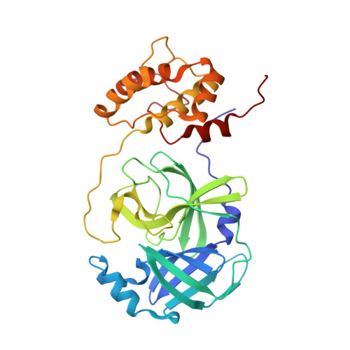 | |
UniProt | |||||
Entity Groups | |||||
| Sequence Clusters | 30% Identity50% Identity70% Identity90% Identity95% Identity100% Identity | ||||
| UniProt Group | K9N7C7 | ||||
Sequence AnnotationsExpand | |||||
Reference Sequence | |||||
| Ligands 2 Unique | |||||
|---|---|---|---|---|---|
| ID | Chains | Name / Formula / InChI Key | 2D Diagram | 3D Interactions | |
| GOL (Subject of Investigation/LOI) Download:Ideal Coordinates CCD File | E [auth A] F [auth A] G [auth A] H [auth B] L [auth C] | GLYCEROL C3 H8 O3 PEDCQBHIVMGVHV-UHFFFAOYSA-N |  | ||
| NA (Subject of Investigation/LOI) Download:Ideal Coordinates CCD File | I [auth B], J [auth B], K [auth B], Q [auth C], R [auth D] | SODIUM ION Na FKNQFGJONOIPTF-UHFFFAOYSA-N |  | ||
| Length ( Å ) | Angle ( ˚ ) |
|---|---|
| a = 130.411 | α = 90 |
| b = 90.01 | β = 108.328 |
| c = 120.517 | γ = 90 |
| Software Name | Purpose |
|---|---|
| CrysalisPro | data collection |
| PHENIX | refinement |
| CrysalisPro | data reduction |
| CrysalisPro | data scaling |
| PHASER | phasing |
| Funding Organization | Location | Grant Number |
|---|---|---|
| Other private | United States | -- |


