8E5G | pdb_00008e5g

Cryo-EM of A. veneficus cytochrome filament


Experimental Data Snapshot

  • Method: ELECTRON MICROSCOPY
  • Resolution: 3.90 Å
  • Aggregation State: FILAMENT 
  • Reconstruction Method: HELICAL 

wwPDB Validation 3D Report Full Report

Validation slider image for 8E5G

This is version 1.2 of the entry. See complete history

Literature

Extracellular cytochrome nanowires appear to be ubiquitous in prokaryotes.

Baquero, D.P.Cvirkaite-Krupovic, V.Hu, S.S.Fields, J.L.Liu, X.Rensing, C.Egelman, E.H.Krupovic, M.Wang, F.

(2023) Cell 186: 2853-2864.e8

  • DOI: https://doi.org/10.1016/j.cell.2023.05.012
  • Primary Citation Related Structures: 
    8E5F, 8E5G

  • PubMed Abstract: 

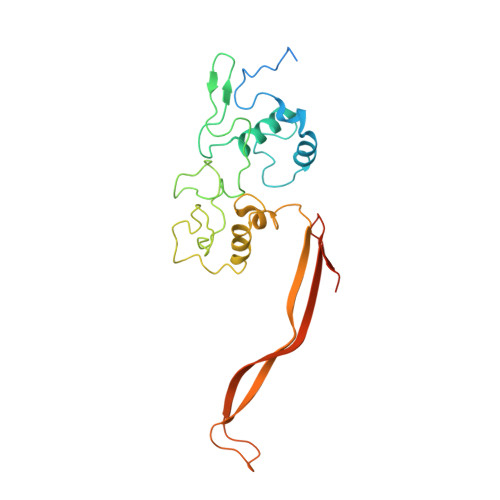
    Electrically conductive appendages from the anaerobic bacterium Geobacter sulfurreducens, recently identified as extracellular cytochrome nanowires (ECNs), have received wide attention due to numerous potential applications. However, whether other organisms employ similar ECNs for electron transfer remains unknown. Here, using cryoelectron microscopy, we describe the atomic structures of two ECNs from two major orders of hyperthermophilic archaea present in deep-sea hydrothermal vents and terrestrial hot springs. Homologs of Archaeoglobus veneficus ECN are widespread among mesophilic methane-oxidizing Methanoperedenaceae, alkane-degrading Syntrophoarchaeales archaea, and in the recently described megaplasmids called Borgs. The ECN protein subunits lack similarities in their folds; however, they share a common heme arrangement, suggesting an evolutionarily optimized heme packing for efficient electron transfer. The detection of ECNs in archaea suggests that filaments containing closely stacked hemes may be a common and widespread mechanism for long-range electron transfer in both prokaryotic domains of life.


  • Organizational Affiliation
    • Institut Pasteur, Université Paris Cité, CNRS UMR6047, Archaeal Virology Unit, Paris 75015, France.

Macromolecule Content 

  • Total Structure Weight: 30.43 kDa 
  • Atom Count: 1,813 
  • Modeled Residue Count: 220 
  • Deposited Residue Count: 262 
  • Unique protein chains: 1

Macromolecules

Find similar proteins by:|  3D Structure
Entity ID: 1
MoleculeChains  Sequence LengthOrganismDetailsImage
c-type cytochrome262Archaeoglobus veneficusMutation(s): 0 
UniProt
Find proteins for F2KMU8 (Archaeoglobus veneficus (strain DSM 11195 / SNP6))
Explore F2KMU8 
Go to UniProtKB:  F2KMU8
Entity Groups
Sequence Clusters30% Identity50% Identity70% Identity90% Identity95% Identity100% Identity
UniProt GroupF2KMU8
Sequence Annotations
Expand
Reference Sequence

Experimental Data & Validation

Experimental Data

  • Method: ELECTRON MICROSCOPY
  • Resolution: 3.90 Å
  • Aggregation State: FILAMENT 
  • Reconstruction Method: HELICAL 

Structure Validation

View Full Validation Report


Entry History 

& Funding Information

Deposition Data


Funding OrganizationLocationGrant Number
National Institutes of Health/National Institute of General Medical Sciences (NIH/NIGMS)United States5K99GM138756-02
National Institutes of Health/National Institute of General Medical Sciences (NIH/NIGMS)United StatesR35GM122510

Revision History  (Full details and data files)

  • Version 1.0: 2023-05-10
    Type: Initial release
  • Version 1.1: 2023-07-26
    Changes: Database references
  • Version 1.2: 2024-10-23
    Changes: Data collection, Structure summary