8E3P | pdb_00008e3p

Co-crystal structure of Chaetomium glucosidase with compound 5


Experimental Data Snapshot

  • Method: X-RAY DIFFRACTION
  • Resolution: 2.30 Å
  • R-Value Free: 
    0.243 (Depositor), 0.243 (DCC) 
  • R-Value Work: 
    0.195 (Depositor), 0.195 (DCC) 
  • R-Value Observed: 
    0.198 (Depositor) 

Starting Model: experimental
View more details

wwPDB Validation 3D Report Full Report

Validation slider image for 8E3P

Ligand Structure Quality Assessment 


This is version 1.3 of the entry. See complete history

Literature

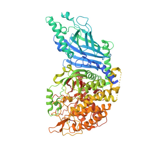
Structure-Based Design of Potent Iminosugar Inhibitors of Endoplasmic Reticulum alpha-Glucosidase I with Anti-SARS-CoV-2 Activity.

Karade, S.S.Franco, E.J.Rojas, A.C.Hanrahan, K.C.Kolesnikov, A.Yu, W.MacKerell Jr., A.D.Hill, D.C.Weber, D.J.Brown, A.N.Treston, A.M.Mariuzza, R.A.

(2023) J Med Chem 66: 2744-2760

  • DOI: https://doi.org/10.1021/acs.jmedchem.2c01750
  • Primary Citation Related Structures: 
    7R6J, 7RD2, 7REV, 8E3J, 8E3P, 8E4I, 8E4K, 8E4Z, 8E5U, 8E6G, 8ECW, 8EGV, 8EHP, 8EID, 8EKN, 8ELE, 8EPJ, 8EPO, 8EPR, 8EQ7, 8EQX, 8ER4, 8ETL, 8ETO, 8EUD, 8EUR, 8EUT, 8EUX

  • PubMed Abstract: 

    Enveloped viruses depend on the host endoplasmic reticulum (ER) quality control (QC) machinery for proper glycoprotein folding. The endoplasmic reticulum quality control (ERQC) enzyme α-glucosidase I (α-GluI) is an attractive target for developing broad-spectrum antivirals. We synthesized 28 inhibitors designed to interact with all four subsites of the α-GluI active site. These inhibitors are derivatives of the iminosugars 1-deoxynojirimycin (1-DNJ) and valiolamine. Crystal structures of ER α-GluI bound to 25 1-DNJ and three valiolamine derivatives revealed the basis for inhibitory potency. We established the structure-activity relationship (SAR) and used the Site Identification by Ligand Competitive Saturation (SILCS) method to develop a model for predicting α-GluI inhibition. We screened the compounds against SARS-CoV-2 in vitro to identify those with greater antiviral activity than the benchmark α-glucosidase inhibitor UV-4. These host-targeting compounds are candidates for investigation in animal models of SARS-CoV-2 and for testing against other viruses that rely on ERQC for correct glycoprotein folding.


  • Organizational Affiliation
    • University of Maryland Institute for Bioscience and Biotechnology Research, Rockville, Maryland 20850, United States.

Macromolecule Content 

  • Total Structure Weight: 189.02 kDa 
  • Atom Count: 12,647 
  • Modeled Residue Count: 1,531 
  • Deposited Residue Count: 1,638 
  • Unique protein chains: 1

Macromolecules

Find similar proteins by:|  3D Structure
Entity ID: 1
MoleculeChains  Sequence LengthOrganismDetailsImage
Chaetomium alpha glucosidase
A, B
819Thermochaetoides thermophila DSM 1495Mutation(s): 0 
Gene Names: CTHT_0061620
EC: 3.2.1.106
UniProt
Find proteins for G0SFD1 (Chaetomium thermophilum (strain DSM 1495 / CBS 144.50 / IMI 039719))
Explore G0SFD1 
Go to UniProtKB:  G0SFD1
Entity Groups
Sequence Clusters30% Identity50% Identity70% Identity90% Identity95% Identity100% Identity
UniProt GroupG0SFD1
Glycosylation
Glycosylation Sites: 1
Sequence Annotations
Expand
Reference Sequence

Small Molecules

Ligands 4 Unique
IDChains Name / Formula / InChI Key2D Diagram3D Interactions
UF6
(Subject of Investigation/LOI)

Query on UF6


Download:Ideal Coordinates CCD File
C [auth A],
J [auth B]
(2S,3S,4S,5R)-2-(hydroxymethyl)-1-{6-[3-nitro-5-(pyridin-4-yl)anilino]hexyl}piperidine-3,4,5-triol
C23 H32 N4 O6
MVGXUAWNTZOMGP-BJESRGMDSA-N
NAG

Query on NAG


Download:Ideal Coordinates CCD File
K [auth B]2-acetamido-2-deoxy-beta-D-glucopyranose
C8 H15 N O6
OVRNDRQMDRJTHS-FMDGEEDCSA-N
SO4

Query on SO4


Download:Ideal Coordinates CCD File
E [auth A]
F [auth A]
G [auth A]
H [auth A]
I [auth A]
E [auth A],
F [auth A],
G [auth A],
H [auth A],
I [auth A],
M [auth B],
N [auth B],
O [auth B],
P [auth B],
Q [auth B],
R [auth B]
SULFATE ION
O4 S
QAOWNCQODCNURD-UHFFFAOYSA-L
GOL

Query on GOL


Download:Ideal Coordinates CCD File
D [auth A],
L [auth B]
GLYCEROL
C3 H8 O3
PEDCQBHIVMGVHV-UHFFFAOYSA-N

Experimental Data & Validation

Experimental Data

  • Method: X-RAY DIFFRACTION
  • Resolution: 2.30 Å
  • R-Value Free:  0.243 (Depositor), 0.243 (DCC) 
  • R-Value Work:  0.195 (Depositor), 0.195 (DCC) 
  • R-Value Observed: 0.198 (Depositor) 
Space Group: I 2 2 2
Unit Cell:
Length ( Å )Angle ( ˚ )
a = 137.353α = 90
b = 178.943β = 90
c = 180.474γ = 90
Software Package:
Software NamePurpose
REFMACrefinement
PDB_EXTRACTdata extraction
HKL-2000data reduction
HKL-2000data scaling
PHASERphasing

Structure Validation

View Full Validation Report


Ligand Structure Quality Assessment 


Entry History 

& Funding Information

Deposition Data


Funding OrganizationLocationGrant Number
Not funded--

Revision History  (Full details and data files)

  • Version 1.0: 2023-02-22
    Type: Initial release
  • Version 1.1: 2023-03-08
    Changes: Database references
  • Version 1.2: 2023-10-25
    Changes: Data collection, Refinement description
  • Version 1.3: 2024-11-06
    Changes: Structure summary