Structure-guided approach to modulate small molecule binding to a promiscuous ligand-activated protein.
Lin, W., Huber, A.D., Poudel, S., Li, Y., Seetharaman, J., Miller, D.J., Chen, T.(2023) Proc Natl Acad Sci U S A 120: e2217804120-e2217804120
- PubMed: 36848571
- DOI: https://doi.org/10.1073/pnas.2217804120
- Primary Citation of Related Structures:
8E3N, 8EQZ, 8FPE - PubMed Abstract:
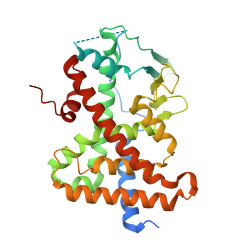
Ligand-binding promiscuity in detoxification systems protects the body from toxicological harm but is a roadblock to drug development due to the difficulty in optimizing small molecules to both retain target potency and avoid metabolic events. Immense effort is invested in evaluating metabolism of molecules to develop safer, more effective treatments, but engineering specificity into or out of promiscuous proteins and their ligands is a challenging task. To better understand the promiscuous nature of detoxification networks, we have used X-ray crystallography to characterize a structural feature of pregnane X receptor (PXR), a nuclear receptor that is activated by diverse molecules (with different structures and sizes) to up-regulate transcription of drug metabolism genes. We found that large ligands expand PXR's ligand-binding pocket, and the ligand-induced expansion occurs through a specific unfavorable compound-protein clash that likely contributes to reduced binding affinity. Removing the clash by compound modification resulted in more favorable binding modes with significantly enhanced binding affinity. We then engineered the unfavorable ligand-protein clash into a potent, small PXR ligand, resulting in marked reduction in PXR binding and activation. Structural analysis showed that PXR is remodeled, and the modified ligands reposition in the binding pocket to avoid clashes, but the conformational changes result in less favorable binding modes. Thus, ligand-induced binding pocket expansion increases ligand-binding potential of PXR but is an unfavorable event; therefore, drug candidates can be engineered to expand PXR's ligand-binding pocket and reduce their safety liability due to PXR binding.
Organizational Affiliation:
Department of Chemical Biology and Therapeutics, St. Jude Children's Research Hospital, Memphis, TN 38105.


