8E37 | pdb_00008e37

Structure of Campylobacter concisus wild-type SeMet PglC


Experimental Data Snapshot

  • Method: X-RAY DIFFRACTION
  • Resolution: 3.01 Å
  • R-Value Free: 
    0.296 (Depositor), 0.296 (DCC) 
  • R-Value Work: 
    0.266 (Depositor), 0.266 (DCC) 
  • R-Value Observed: 
    0.267 (Depositor) 

Starting Model: experimental
View more details

wwPDB Validation 3D Report Full Report

Validation slider image for 8E37

This is version 1.4 of the entry. See complete history

Literature

Synergistic computational and experimental studies of a phosphoglycosyl transferase membrane/ligand ensemble.

Majumder, A.Vuksanovic, N.Ray, L.C.Bernstein, H.M.Allen, K.N.Imperiali, B.Straub, J.E.

(2023) J Biological Chem 299: 105194-105194

  • DOI: https://doi.org/10.1016/j.jbc.2023.105194
  • Primary Citation Related Structures: 
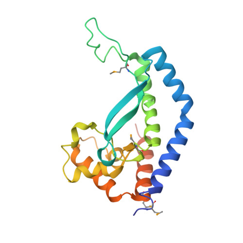
    8E37

  • PubMed Abstract: 

    Complex glycans serve essential functions in all living systems. Many of these intricate and byzantine biomolecules are assembled employing biosynthetic pathways wherein the constituent enzymes are membrane-associated. A signature feature of the stepwise assembly processes is the essentiality of unusual linear long-chain polyprenol phosphate-linked substrates of specific isoprene unit geometry, such as undecaprenol phosphate (UndP) in bacteria. How these enzymes and substrates interact within a lipid bilayer needs further investigation. Here, we focus on a small enzyme, PglC from Campylobacter, structurally characterized for the first time in 2018 as a detergent-solubilized construct. PglC is a monotopic phosphoglycosyl transferase that embodies the functional core structure of the entire enzyme superfamily and catalyzes the first membrane-committed step in a glycoprotein assembly pathway. The size of the enzyme is significant as it enables high-level computation and relatively facile, for a membrane protein, experimental analysis. Our ensemble computational and experimental results provided a high-level view of the membrane-embedded PglC/UndP complex. The findings suggested that it is advantageous for the polyprenol phosphate to adopt a conformation in the same leaflet where the monotopic membrane protein resides as opposed to additionally disrupting the opposing leaflet of the bilayer. Further, the analysis showed that electrostatic steering acts as a major driving force contributing to the recognition and binding of both UndP and the soluble nucleotide sugar substrate. Iterative computational and experimental mutagenesis support a specific interaction of UndP with phosphoglycosyl transferase cationic residues and suggest a role for critical conformational transitions in substrate binding and specificity.


  • Organizational Affiliation
    • Department of Chemistry, Boston University, Boston, Massachusetts, USA.

Macromolecule Content 

  • Total Structure Weight: 188.94 kDa 
  • Atom Count: 12,088 
  • Modeled Residue Count: 1,480 
  • Deposited Residue Count: 1,640 
  • Unique protein chains: 1

Macromolecules

Find similar proteins by:|  3D Structure
Entity ID: 1
MoleculeChains  Sequence LengthOrganismDetailsImage
N,N'-diacetylbacilliosaminyl-1-phosphate transferase205Campylobacter concisus 13826Mutation(s): 0 
Gene Names: pglCCCC13826_0450
EC: 2.7.8.36
UniProt
Find proteins for A7ZET4 (Campylobacter concisus (strain 13826))
Explore A7ZET4 
Go to UniProtKB:  A7ZET4
Entity Groups
Sequence Clusters30% Identity50% Identity70% Identity90% Identity95% Identity100% Identity
UniProt GroupA7ZET4
Sequence Annotations
Expand
Reference Sequence

Small Molecules

Modified Residues  1 Unique
IDChains TypeFormula2D DiagramParent
MSE
Query on MSE
A [auth B]
B [auth A]
C
D
E
A [auth B],
B [auth A],
C,
D,
E,
F,
G,
H
L-PEPTIDE LINKINGC5 H11 N O2 SeMET

Experimental Data & Validation

Experimental Data

  • Method: X-RAY DIFFRACTION
  • Resolution: 3.01 Å
  • R-Value Free:  0.296 (Depositor), 0.296 (DCC) 
  • R-Value Work:  0.266 (Depositor), 0.266 (DCC) 
  • R-Value Observed: 0.267 (Depositor) 
Space Group: P 31 2 1
Unit Cell:
Length ( Å )Angle ( ˚ )
a = 142.82α = 90
b = 142.82β = 90
c = 192.563γ = 120
Software Package:
Software NamePurpose
PHENIXrefinement
XDSdata reduction
XDSdata scaling
PHASERphasing

Structure Validation

View Full Validation Report


Entry History 

& Funding Information

Deposition Data


Funding OrganizationLocationGrant Number
National Institutes of Health/National Institute of General Medical Sciences (NIH/NIGMS)United States2R01GM131627

Revision History  (Full details and data files)

  • Version 1.0: 2023-09-06
    Type: Initial release
  • Version 1.1: 2023-10-04
    Changes: Database references
  • Version 1.2: 2023-11-15
    Changes: Data collection
  • Version 1.3: 2024-10-09
    Changes: Structure summary
  • Version 1.4: 2026-03-04
    Changes: Refinement description