Cryo-EM Structure of the Type IV Pilus Extension ATPase from Enteropathogenic Escherichia coli.
Nayak, A.R., Singh, P.K., Zhao, J., Samso, M., Donnenberg, M.S.(2022) mBio 13: e0227022-e0227022
- PubMed: 36326250 Search on PubMedSearch on PubMed Central
- DOI: https://doi.org/10.1128/mbio.02270-22
- Primary Citation Related Structures:
8DZE, 8DZF, 8DZG - PubMed Abstract:
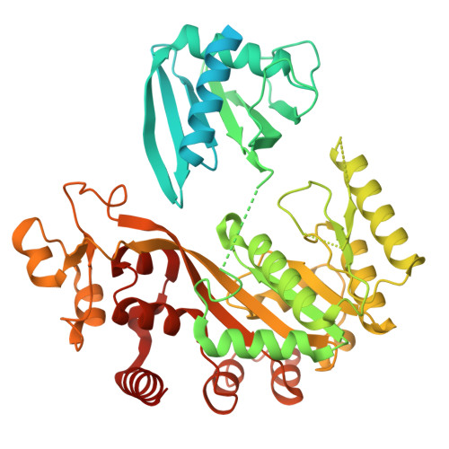
Type 4 pili (T4P) are retractable surface appendages found on numerous bacteria and archaea that play essential roles in various microbial functions, including host colonization by pathogens. An ATPase is required for T4P extension, but the mechanism by which chemical energy is transduced to mechanical energy for pilus extension has not been elucidated. Here, we report the cryo-electron microscopy (cryo-EM) structure of the BfpD ATPase from enteropathogenic Escherichia coli (EPEC) in the presence of either ADP or a mixture of ADP and AMP-PNP. Both structures, solved at 3 Å resolution, reveal the typical toroid shape of AAA+ ATPases and unambiguous 6-fold symmetry. This 6-fold symmetry contrasts with the 2-fold symmetry previously reported for other T4P extension ATPase structures, all of which were from thermophiles and solved by crystallography. In the presence of the nucleotide mixture, BfpD bound exclusively AMP-PNP, and this binding resulted in a modest outward expansion in comparison to the structure in the presence of ADP, suggesting a concerted model for hydrolysis. De novo molecular models reveal a partially open configuration of all subunits where the nucleotide binding site may not be optimally positioned for catalysis. ATPase functional studies reveal modest activity similar to that of other extension ATPases, while calculations indicate that this activity is insufficient to power pilus extension. Our results reveal that, despite similarities in primary sequence and tertiary structure, T4P extension ATPases exhibit divergent quaternary configurations. Our data raise new possibilities regarding the mechanism by which T4P extension ATPases power pilus formation. IMPORTANCE Type 4 pili are hairlike surface appendages on many bacteria and archaea that can be extended and retracted with tremendous force. They play a critical role in disease caused by several deadly human pathogens. Pilus extension is made possible by an enzyme that converts chemical energy to mechanical energy. Here, we describe the three-dimensional structure of such an enzyme from a human pathogen in unprecedented detail, which reveals a mechanism of action that has not been seen previously among enzymes that power type 4 pilus extension.
- Department of Physiology and Biophysics, Virginia Commonwealth Universitygrid.224260.0, Richmond, Virginia, USA.
Organizational Affiliation: