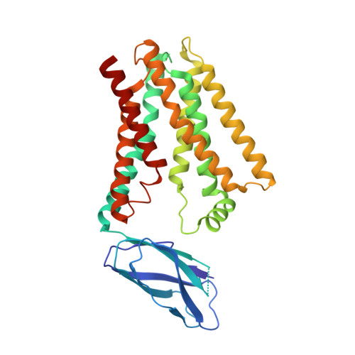
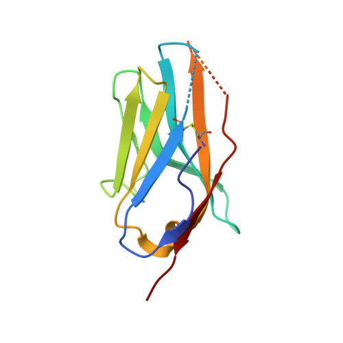
8DYP

Crystal structure of human cystine transporter cystinosin


Experimental Data Snapshot

  • Method: X-RAY DIFFRACTION
  • Resolution: 3.40 Å
  • R-Value Free: 0.302 
  • R-Value Work: 0.270 
  • R-Value Observed: 0.272 

Starting Models: experimental
View more details

wwPDB Validation   3D Report Full Report


Ligand Structure Quality Assessment 


This is version 1.2 of the entry. See complete history


Literature

Structure and mechanism of human cystine exporter cystinosin.

Guo, X.Schmiege, P.Assafa, T.E.Wang, R.Xu, Y.Donnelly, L.Fine, M.Ni, X.Jiang, J.Millhauser, G.Feng, L.Li, X.

(2022) Cell 185: 3739-3752.e18

  • DOI: https://doi.org/10.1016/j.cell.2022.08.020
  • Primary Citation of Related Structures:  
    8DKE, 8DKI, 8DKM, 8DKW, 8DKX, 8DYP

  • PubMed Abstract: 

    Lysosomal amino acid efflux by proton-driven transporters is essential for lysosomal homeostasis, amino acid recycling, mTOR signaling, and maintaining lysosomal pH. To unravel the mechanisms of these transporters, we focus on cystinosin, a prototypical lysosomal amino acid transporter that exports cystine to the cytosol, where its reduction to cysteine supplies this limiting amino acid for diverse fundamental processes and controlling nutrient adaptation. Cystinosin mutations cause cystinosis, a devastating lysosomal storage disease. Here, we present structures of human cystinosin in lumen-open, cytosol-open, and cystine-bound states, which uncover the cystine recognition mechanism and capture the key conformational states of the transport cycle. Our structures, along with functional studies and double electron-electron resonance spectroscopic investigations, reveal the molecular basis for the transporter's conformational transitions and protonation switch, show conformation-dependent Ragulator-Rag complex engagement, and demonstrate an unexpected activation mechanism. These findings provide molecular insights into lysosomal amino acid efflux and a potential therapeutic strategy.


  • Organizational Affiliation

    Department of Molecular and Cellular Physiology, Stanford University School of Medicine, Stanford, CA 94305, USA.


Macromolecules
Find similar proteins by:  (by identity cutoff)  |  3D Structure
Entity ID: 1
MoleculeChains Sequence LengthOrganismDetailsImage
Cystinosin332Homo sapiensMutation(s): 1 
Gene Names: CTNS
Membrane Entity: Yes 
UniProt & NIH Common Fund Data Resources
Find proteins for O60931 (Homo sapiens)
Explore O60931 
Go to UniProtKB:  O60931
PHAROS:  O60931
GTEx:  ENSG00000040531 
Entity Groups  
Sequence Clusters30% Identity50% Identity70% Identity90% Identity95% Identity100% Identity
UniProt GroupO60931
Sequence Annotations
Expand
  • Reference Sequence
Find similar proteins by:  (by identity cutoff)  |  3D Structure
Entity ID: 2
MoleculeChains Sequence LengthOrganismDetailsImage
Nanobody P10118synthetic constructMutation(s): 0 
Entity Groups  
Sequence Clusters30% Identity50% Identity70% Identity90% Identity95% Identity100% Identity
Sequence Annotations
Expand
  • Reference Sequence
Experimental Data & Validation

Experimental Data

  • Method: X-RAY DIFFRACTION
  • Resolution: 3.40 Å
  • R-Value Free: 0.302 
  • R-Value Work: 0.270 
  • R-Value Observed: 0.272 
  • Space Group: P 2 21 21
Unit Cell:
Length ( Å )Angle ( ˚ )
a = 49.51α = 90
b = 71.27β = 90
c = 194.64γ = 90
Software Package:
Software NamePurpose
HKL-2000data scaling
HKL-2000data reduction
PHENIXrefinement
PHASERphasing

Structure Validation

View Full Validation Report


Ligand Structure Quality Assessment 


Entry History & Funding Information

Deposition Data


Funding OrganizationLocationGrant Number
National Institutes of Health/National Institute of General Medical Sciences (NIH/NIGMS)United States--

Revision History  (Full details and data files)

  • Version 1.0: 2022-11-16
    Type: Initial release
  • Version 1.1: 2023-10-18
    Changes: Data collection, Refinement description
  • Version 1.2: 2024-10-23
    Changes: Structure summary