8DT3 | pdb_00008dt3

Cryo-EM structure of spike binding to Fab of neutralizing antibody (locally refined)


Experimental Data Snapshot

  • Method: ELECTRON MICROSCOPY
  • Resolution: 3.30 Å
  • Aggregation State: PARTICLE 
  • Reconstruction Method: SINGLE PARTICLE 

wwPDB Validation 3D Report Full Report

Validation slider image for 8DT3

This is version 1.3 of the entry. See complete history

Literature

An antibody that neutralizes SARS-CoV-1 and SARS-CoV-2 by binding to a conserved spike epitope outside the receptor binding motif.

Fang, Y.Sun, P.Xie, X.Du, M.Du, F.Ye, J.Kalveram, B.K.Plante, J.A.Plante, K.S.Li, B.Bai, X.C.Shi, P.Y.Chen, Z.J.

(2022) Sci Immunol 7: eabp9962-eabp9962

  • DOI: https://doi.org/10.1126/sciimmunol.abp9962
  • Primary Citation Related Structures: 
    8DT3

  • PubMed Abstract: 

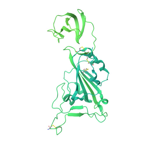
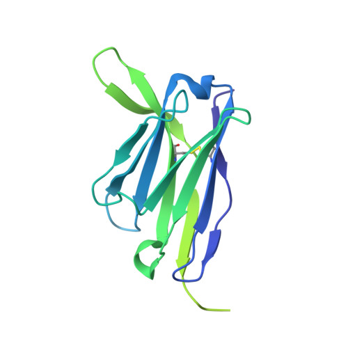
    The rapid evolution of severe acute respiratory syndrome coronavirus 2 (SARS-CoV-2), such as the Omicron variants that are highly transmissible and immune evasive, underscores the need to develop therapeutic antibodies with broad neutralizing activities. Here, we used the LIBRA-seq technology, which identified SARS-CoV-2-specific B cells via DNA barcoding and subsequently single-cell sequenced BCRs, to identify an antibody, SW186, which could neutralize major SARS-CoV-2 variants of concern, including Beta, Delta, and Omicron, as well as SARS-CoV-1. The cryo-EM structure of SW186 bound to the receptor binding domain (RBD) of the viral spike protein showed that SW186 interacted with an epitope of the RBD that is not at the interface of its binding to the ACE2 receptor but is highly conserved among SARS coronaviruses. This epitope encompasses a glycosylation site (N343) of the viral spike protein. Administration of SW186 in mice after they were infected with SARS-CoV-2 Alpha, Beta, or Delta variants reduced the viral loads in the lung. These results demonstrated that SW186 neutralizes diverse SARS coronaviruses by binding to a conserved RBD epitope, which could serve as a target for further antibody development.


  • Organizational Affiliation
    • Department of Molecular Biology, University of Texas Southwestern Medical Center, Dallas, TX 75390-9148, USA.

Macromolecule Content 

  • Total Structure Weight: 192.78 kDa 
  • Atom Count: 3,821 
  • Modeled Residue Count: 485 
  • Deposited Residue Count: 1,744 
  • Unique protein chains: 3

Macromolecules

Find similar proteins by:|  3D Structure
Entity ID: 1
MoleculeChains  Sequence LengthOrganismDetailsImage
Spike glycoproteinA [auth C]1,281Severe acute respiratory syndrome coronavirus 2Mutation(s): 0 
Gene Names: S2
UniProt
Find proteins for P0DTC2 (Severe acute respiratory syndrome coronavirus 2)
Explore P0DTC2 
Go to UniProtKB:  P0DTC2
Entity Groups
Sequence Clusters30% Identity50% Identity70% Identity90% Identity95% Identity100% Identity
UniProt GroupP0DTC2
Glycosylation
Glycosylation Sites: 1Go to GlyGen: P0DTC2-1
Sequence Annotations
Expand
Reference Sequence
Find similar proteins by:|  3D Structure
Entity ID: 2
MoleculeChains  Sequence LengthOrganismDetailsImage
Heavy chain Fab of SW186B [auth H]232Mus musculusMutation(s): 0 
Find similar proteins by:|  3D Structure
Entity ID: 3
MoleculeChains  Sequence LengthOrganismDetailsImage
Light chain Fab of SW186C [auth L]231Mus musculusMutation(s): 0 

Oligosaccharides

Help  
Entity ID: 4
MoleculeChains Length2D Diagram GlycosylationD Interactions
2-acetamido-2-deoxy-beta-D-glucopyranose-(1-4)-[alpha-L-fucopyranose-(1-6)]2-acetamido-2-deoxy-beta-D-glucopyranoseD [auth T]3N-Glycosylation
Glycosylation Resources
GlyTouCan: G21290RB
GlyCosmos: G21290RB
GlyGen: G21290RB

Experimental Data & Validation

Experimental Data

  • Method: ELECTRON MICROSCOPY
  • Resolution: 3.30 Å
  • Aggregation State: PARTICLE 
  • Reconstruction Method: SINGLE PARTICLE 
EM Software:
TaskSoftware PackageVersion
RECONSTRUCTIONRELION
MODEL REFINEMENTPHENIX

Structure Validation

View Full Validation Report


Entry History 

& Funding Information

Deposition Data


Funding OrganizationLocationGrant Number
Howard Hughes Medical Institute (HHMI)United States--

Revision History  (Full details and data files)

  • Version 1.0: 2022-08-03
    Type: Initial release
  • Version 1.1: 2022-08-17
    Changes: Database references
  • Version 1.2: 2022-11-09
    Changes: Database references
  • Version 1.3: 2024-11-06
    Changes: Data collection, Structure summary