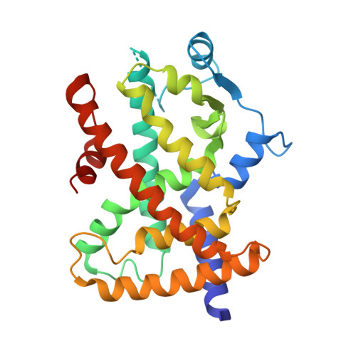
RCSB PDB - 8DSZ: PPARg bound to partial agonist H3B-487

 8DSZ

PPARg bound to partial agonist H3B-487


Experimental Data Snapshot

  • Method: X-RAY DIFFRACTION
  • Resolution: 2.50 Å
  • R-Value Free: 0.269 
  • R-Value Work: 0.208 
  • R-Value Observed: 0.211 

Starting Model: experimental
View more details

wwPDB Validation   3D Report Full Report


Ligand Structure Quality Assessment 

Created with Raphaël 2.3.0Worse 01 BetterLigand structure goodness of fit to experimental dataBest fitted TJOClick on this verticalbar to view details

This is version 1.2 of the entry. See complete history


Literature

Biochemical and structural basis for the pharmacological inhibition of nuclear hormone receptor PPAR gamma by inverse agonists.

Irwin, S.Karr, C.Furman, C.Tsai, J.Gee, P.Banka, D.Wibowo, A.S.Dementiev, A.A.O'Shea, M.Yang, J.Lowe, J.Mitchell, L.Ruppel, S.Fekkes, P.Zhu, P.Korpal, M.Larsen, N.A.

(2022) J Biol Chem 298: 102539-102539

  • DOI: https://doi.org/10.1016/j.jbc.2022.102539
  • Primary Citation of Related Structures:  
    8DKN, 8DKV, 8DSY, 8DSZ

  • PubMed Abstract: 

    Recent studies have reported that the peroxisome proliferator-activated receptor gamma (PPARγ) pathway is activated in approximately 40% of patients with muscle-invasive bladder cancer. This led us to investigate pharmacological repression of PPARγ as a possible intervention strategy. Here, we characterize PPARγ antagonists and inverse agonists and find that the former behave as silent ligands, whereas inverse agonists (T0070907 and SR10221) repress downstream PPARγ target genes leading to growth inhibition in bladder cancer cell lines. To understand the mechanism, we determined the ternary crystal structure of PPARγ bound to T0070907 and the corepressor (co-R) peptide NCOR1. The structure shows that the AF-2 helix 12 (H12) rearranges to bind inside the ligand-binding domain, where it forms stabilizing interactions with the compound. This dramatic movement in H12 unveils a large interface for co-R binding. In contrast, the crystal structure of PPARγ bound to a SR10221 analog shows more subtle structural differences, where the compound binds and pushes H12 away from the ligand-binding domain to allow co-R binding. Interestingly, we found that both classes of compound promote recruitment of co-R proteins in biochemical assays but with distinct conformational changes in H12. We validate our structural models using both site-directed mutagenesis and chemical probes. Our findings offer new mechanistic insights into pharmacological modulation of PPARγ signaling.


  • Organizational Affiliation

    Department of Drug Discovery, H3 Biomedicine, Cambridge, Massachusetts, USA.


Macromolecules
Find similar proteins by:  (by identity cutoff)  |  3D Structure
Entity ID: 1
MoleculeChains Sequence LengthOrganismDetailsImage
Peroxisome proliferator-activated receptor gamma
A, B
273Homo sapiensMutation(s): 0 
Gene Names: PPARGNR1C3
UniProt & NIH Common Fund Data Resources
Find proteins for P37231 (Homo sapiens)
Explore P37231 
Go to UniProtKB:  P37231
PHAROS:  P37231
GTEx:  ENSG00000132170 
Entity Groups  
Sequence Clusters30% Identity50% Identity70% Identity90% Identity95% Identity100% Identity
UniProt GroupP37231
Sequence Annotations
Expand
  • Reference Sequence
Small Molecules
Ligands 1 Unique
IDChains Name / Formula / InChI Key2D Diagram3D Interactions
TJO (Subject of Investigation/LOI)
Query on TJO

Download Ideal Coordinates CCD File 
C [auth A],
D [auth B]
(2R)-2-{5-[(5-{[(1R)-1-(4-tert-butylphenyl)ethyl]carbamoyl}-2,3-dimethyl-1H-indol-1-yl)methyl]-2-chlorophenoxy}propanoic acid
C33 H37 Cl N2 O4
NPMSSJHJPGMEJW-IFMALSPDSA-N
Experimental Data & Validation

Experimental Data

  • Method: X-RAY DIFFRACTION
  • Resolution: 2.50 Å
  • R-Value Free: 0.269 
  • R-Value Work: 0.208 
  • R-Value Observed: 0.211 
  • Space Group: C 1 2 1
Unit Cell:
Length ( Å )Angle ( ˚ )
a = 92.975α = 90
b = 60.953β = 103.53
c = 118.842γ = 90
Software Package:
Software NamePurpose
REFMACrefinement
SCALEPACKdata scaling
PDB_EXTRACTdata extraction
HKL-2000data reduction
MOLREPphasing

Structure Validation

View Full Validation Report


Ligand Structure Quality Assessment 

Created with Raphaël 2.3.0Worse 01 BetterLigand structure goodness of fit to experimental dataBest fitted TJOClick on this verticalbar to view details

Entry History & Funding Information

Deposition Data

  • Released Date: 2022-09-07 
  • Deposition Author(s): Larsen, N.A.

Funding OrganizationLocationGrant Number
Other privateUnited States--

Revision History  (Full details and data files)

  • Version 1.0: 2022-09-07
    Type: Initial release
  • Version 1.1: 2023-04-05
    Changes: Database references
  • Version 1.2: 2023-10-25
    Changes: Data collection, Refinement description