8DSK | pdb_00008dsk

Structure of the N358Y variant of serine hydroxymethyltransferase 8 in complex with PLP, glycine, and formyl tetrahydrofolate


Experimental Data Snapshot

  • Method: X-RAY DIFFRACTION
  • Resolution: 1.63 Å
  • R-Value Free: 
    0.214 (Depositor), 0.213 (DCC) 
  • R-Value Work: 
    0.184 (Depositor), 0.184 (DCC) 
  • R-Value Observed: 
    0.186 (Depositor) 

Starting Model: experimental
View more details

wwPDB Validation 3D Report Full Report

Validation slider image for 8DSK

Ligand Structure Quality Assessment 


This is version 1.2 of the entry. See complete history

Literature

Structural and functional analysis of two SHMT8 variants associated with soybean cyst nematode resistance.

Korasick, D.A.Owuocha, L.F.Kandoth, P.K.Tanner, J.J.Mitchum, M.G.Beamer, L.J.

(2024) FEBS J 291: 323-337

  • DOI: https://doi.org/10.1111/febs.16971
  • Primary Citation Related Structures: 
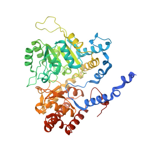
    7UJH, 7UJI, 8DOM, 8DSK, 8FSD

  • PubMed Abstract: 

    Two amino acid variants in soybean serine hydroxymethyltransferase 8 (SHMT8) are associated with resistance to the soybean cyst nematode (SCN), a devastating agricultural pathogen with worldwide economic impacts on soybean production. SHMT8 is a cytoplasmic enzyme that catalyzes the pyridoxal 5-phosphate-dependent conversion of serine and tetrahydrofolate (THF) to glycine and 5,10-methylenetetrahydrofolate. A previous study of the P130R/N358Y double variant of SHMT8, identified in the SCN-resistant soybean cultivar (cv.) Forrest, showed profound impairment of folate binding affinity and reduced THF-dependent enzyme activity, relative to the highly active SHMT8 in cv. Essex, which is susceptible to SCN. Given the importance of SCN-resistance in soybean agriculture, we report here the biochemical and structural characterization of the P130R and N358Y single variants to elucidate their individual effects on soybean SHMT8. We find that both single variants have reduced THF-dependent catalytic activity relative to Essex SHMT8 (10- to 50-fold decrease in k cat /K m ) but are significantly more active than the P130R/N368Y double variant. The kinetic data also show that the single variants lack THF-substrate inhibition as found in Essex SHMT8, an observation with implications for regulation of the folate cycle. Five crystal structures of the P130R and N358Y variants in complex with various ligands (resolutions from 1.49 to 2.30 Å) reveal distinct structural impacts of the mutations and provide new insights into allosterism. Our results support the notion that the P130R/N358Y double variant in Forrest SHMT8 produces unique and unexpected effects on the enzyme, which cannot be easily predicted from the behavior of the individual variants.


  • Organizational Affiliation
    • Department of Biochemistry, University of Missouri, Columbia, MO, USA.

Macromolecule Content 

  • Total Structure Weight: 220.44 kDa 
  • Atom Count: 17,198 
  • Modeled Residue Count: 1,886 
  • Deposited Residue Count: 1,964 
  • Unique protein chains: 1

Macromolecules

Find similar proteins by:|  3D Structure
Entity ID: 1
MoleculeChains  Sequence LengthOrganismDetailsImage
Serine hydroxymethyltransferase
A, B, C, D
491Glycine maxMutation(s): 1 
Gene Names: 100305380GLYMA_08G108900
EC: 2.1.2.1
UniProt
Find proteins for A0A0R0IK90 (Glycine max)
Explore A0A0R0IK90 
Go to UniProtKB:  A0A0R0IK90
Entity Groups
Sequence Clusters30% Identity50% Identity70% Identity90% Identity95% Identity100% Identity
UniProt GroupA0A0R0IK90
Sequence Annotations
Expand
Reference Sequence

Small Molecules

Ligands 3 Unique
IDChains Name / Formula / InChI Key2D Diagram3D Interactions
FFO
(Subject of Investigation/LOI)

Query on FFO


Download:Ideal Coordinates CCD File
F [auth A],
O [auth B],
U [auth C],
Z [auth D]
N-[4-({[(6S)-2-amino-5-formyl-4-oxo-3,4,5,6,7,8-hexahydropteridin-6-yl]methyl}amino)benzoyl]-L-glutamic acid
C20 H23 N7 O7
VVIAGPKUTFNRDU-STQMWFEESA-N
PLG
(Subject of Investigation/LOI)

Query on PLG


Download:Ideal Coordinates CCD File
E [auth A],
N [auth B],
T [auth C],
Y [auth D]
N-GLYCINE-[3-HYDROXY-2-METHYL-5-PHOSPHONOOXYMETHYL-PYRIDIN-4-YL-METHANE]
C10 H15 N2 O7 P
FEVQWBMNLWUBTF-UHFFFAOYSA-N
EDO

Query on EDO


Download:Ideal Coordinates CCD File
AA [auth D]
BA [auth D]
CA [auth D]
DA [auth D]
G [auth A]
AA [auth D],
BA [auth D],
CA [auth D],
DA [auth D],
G [auth A],
H [auth A],
I [auth A],
J [auth A],
K [auth A],
L [auth A],
M [auth B],
P [auth B],
Q [auth B],
R [auth B],
S [auth B],
V [auth C],
W [auth C],
X [auth C]
1,2-ETHANEDIOL
C2 H6 O2
LYCAIKOWRPUZTN-UHFFFAOYSA-N

Experimental Data & Validation

Experimental Data

  • Method: X-RAY DIFFRACTION
  • Resolution: 1.63 Å
  • R-Value Free:  0.214 (Depositor), 0.213 (DCC) 
  • R-Value Work:  0.184 (Depositor), 0.184 (DCC) 
  • R-Value Observed: 0.186 (Depositor) 
Space Group: P 1 21 1
Unit Cell:
Length ( Å )Angle ( ˚ )
a = 87.106α = 90
b = 90.798β = 90.551
c = 147.441γ = 90
Software Package:
Software NamePurpose
PHENIXrefinement
XDSdata reduction
Aimlessdata scaling
PHASERphasing

Structure Validation

View Full Validation Report


Ligand Structure Quality Assessment 


Entry History 

& Funding Information

Deposition Data


Funding OrganizationLocationGrant Number
National Science Foundation (NSF, United States)United StatesIOS 2152548

Revision History  (Full details and data files)

  • Version 1.0: 2023-10-18
    Type: Initial release
  • Version 1.1: 2024-01-31
    Changes: Database references
  • Version 1.2: 2026-03-04
    Changes: Refinement description, Structure summary